Solitary fibrous tumor of the pleura: An analysis of forty patients.
J Thorac Dis. 2012 Apr 1;4(2):146-54. doi: 10.3978/j.issn.2072-1439.2012.01.05.
Agarwal VK, Plotkin BE, Dumani D, et al.
Solitary fibrous tumor of pleura: a case report and review of clinical, radiographic and histologic findings.
J Radiol Case Rep. 2009;3(5):16-20. doi: 10.3941/jrcr.v3i5.200. Epub 2009 May 1.
Sekiya M, Yoshimi K, Muraki K, et al.
Solitary fibrous tumor of the pleura: ultrasonographic imaging findings of 3 cases.
Respir Investig. 2013 Sep;51(3):200-4. doi: 10.1016/j.resinv.2013.04.001. Epub 2013 Jun 13.
Moureau-Zabotto L, Chetaille B, et al.
Solitary fibrous tumor of the prostate: case report and review of the literature.
Case Rep Oncol. 2012 Jan;5(1):22-9. doi: 10.1159/000335680. Epub 2012 Jan 10.
Li JP, Xie CM, Zhang R,et al.
[Imaging features and clinicopathological manifestations of solitary fibrous tumors].
Zhonghua Zhong Liu Za Zhi. 2010 May;32(5):363-7. Chinese.
Nomura T, Satoh R, Kashima K, et al.
A case of large solitary fibrous tumor in the retroperitoneum.
Clin Med Case Rep. 2009 Mar 23;2:21-5. eCollection 2009.
Inaoka T, Takahashi K, Miyokawa N, et al.
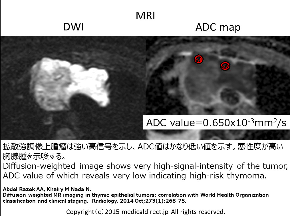
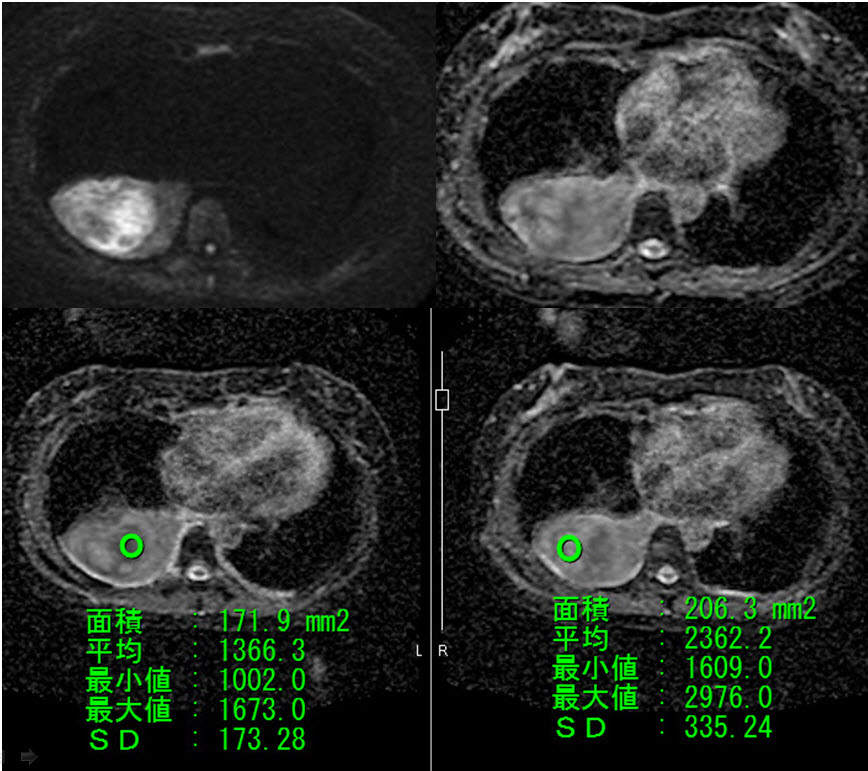
Solitary fibrous tumor of the pleura: apparent diffusion coefficient (ADC) value and ADC map to predict malignant transformation.
J Magn Reson Imaging. 2007 Jul;26(1):155-8.
Aquino SL, Chiles C, Halford P.
Distinction of consolidative bronchioloalveolar carcinoma from pneumonia: do CT criteria work?
AJR Am J Roentgenol. 1998 Aug;171(2):359-63. http://www.ajronline.org/cgi/reprint/171/2/359
Akira M, Atagi S, Kawahara M, Iuchi K, Johkoh T.
High-resolution CT findings of diffuse bronchioloalveolar carcinoma in 38 patients.
AJR Am J Roentgenol. 1999 Dec;173(6):1623-9. http://www.ajronline.org/cgi/reprint/173/6/1623
Kim TH, Kim SJ, Ryu YH, et al.
Differential CT features of infectious pneumonia versus bronchioloalveolar carcinoma (BAC) mimicking pneumonia.
Eur Radiol. 2006 Aug;16(8):1763-8. Epub 2006 Jan 18.
Patsios D, Roberts HC, Paul NS, et al.
Pictorial review of the many faces of bronchioloalveolar cell carcinoma.
Br J Radiol. 2007 Dec;80(960):1015-23. Epub 2007 Oct 16. http://bjr.birjournals.org/cgi/reprint/80/960/1015
Sawada E, Nambu A, Motosugi U, et al.
Localized mucinous bronchioloalveolar carcinoma of the lung: thin-section computed tomography and fluorodeoxyglucose positron emission tomography findings.
Jpn J Radiol. 2010 May;28(4):251-8. Epub 2010 May 29.
CT angiogram sign 関連
Im JG, Han MC, Yu EJ, et al.
Lobar bronchioloalveolar carcinoma: “angiogram sign” on CT scans.
Radiology. 1990 Sep;176(3):749-53.
Vincent JM, Ng YY, Norton AJ, Armstrong P.
CT “angiogram sign” in primary pulmonary lymphoma.
J Comput Assist Tomogr. 1992 Sep-Oct;16(5):829-31.
Murayama S, Onitsuka H, Murakami J, et al.
“CT angiogram sign” in obstructive pneumonitis and pneumonia.
J Comput Assist Tomogr. 1993 Jul-Aug;17(4):609-12.
Shah RM, Friedman AC.
CT angiogram sign: incidence and significance in lobar consolidations evaluated by contrast-enhanced CT.
AJR Am J Roentgenol. 1998 Mar;170(3):719-21. http://www.ajronline.org/cgi/reprint/170/3/719
Maldonado RL.
The CT angiogram sign.
Radiology. 1999 Feb;210(2):323-4.
King LJ, Padley SP, Wotherspoon AC, Nicholson AG.
Pulmonary MALT lymphoma: imaging findings in 24 cases.
Eur Radiol. 2000;10(12):1932-8.
Lee DK, Im JG, Lee KS, Lee JS, Seo JB, Goo JM, Kim TS, Lee JW.
B-cell lymphoma of bronchus-associated lymphoid tissue (BALT): CT features in 10 patients.
J Comput Assist Tomogr. 2000 Jan-Feb;24(1):30-4.
LCH肺
Abbott GF, Rosado-de-Christenson ML, et al.
From the archives of the AFIP: pulmonary Langerhans cell
histiocytosis.
Radiographics. 2004 May-Jun;24(3):821-41.
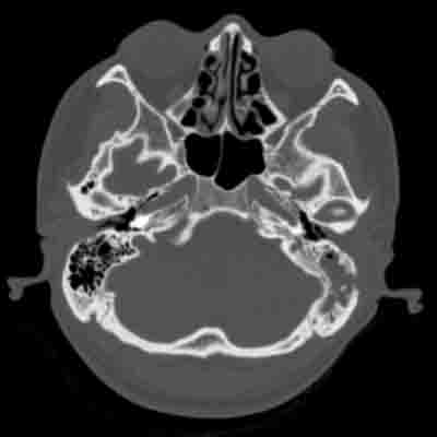
D’Ambrosio N, Soohoo S, Warshall C, Johnson A, Karimi S.
Craniofacial and intracranial manifestations of langerhans
cell histiocytosis: report of findings in 100 patients.
AJR Am J Roentgenol. 2008 Aug;191(2):589-97.

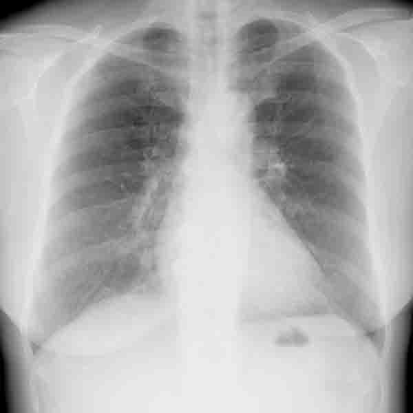
 1 年前正常時胸部単純写真
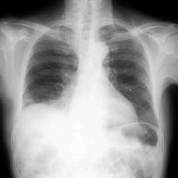 10 ヶ月前肺炎発症時
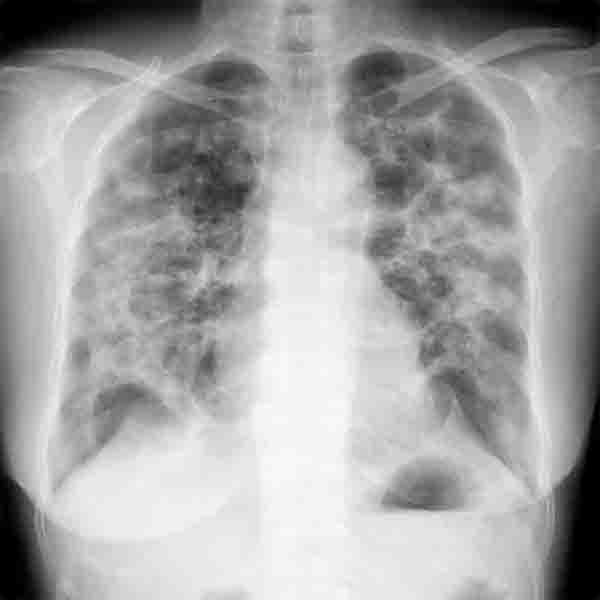
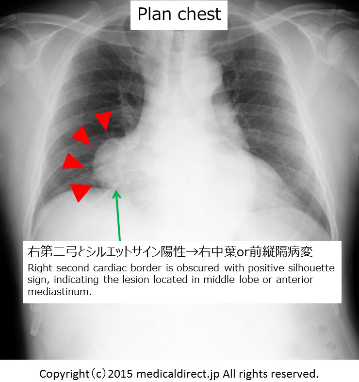
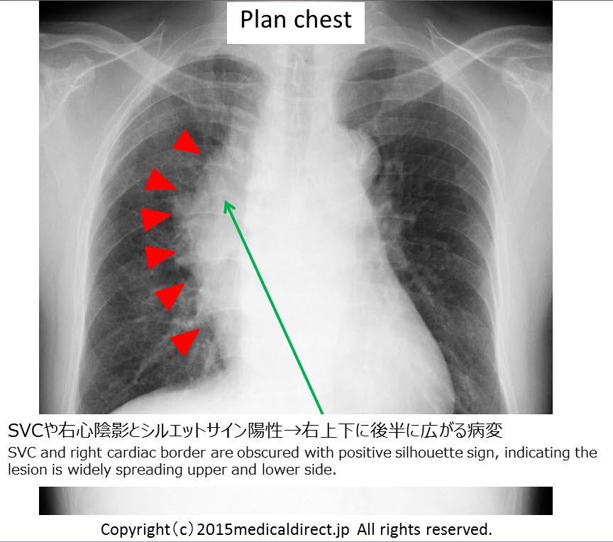
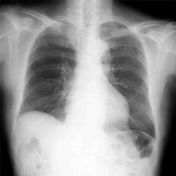
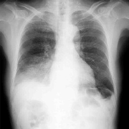 今回の胸部単純写真正面像
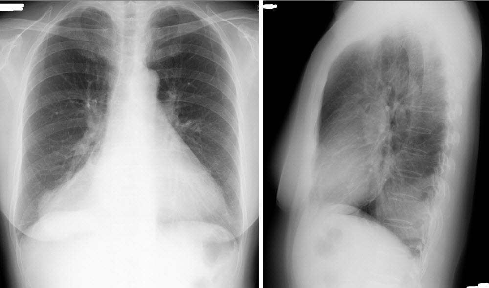
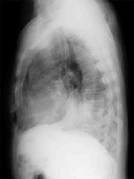 今回の胸部単純写真側面像
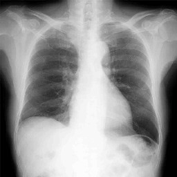 肺炎改善後
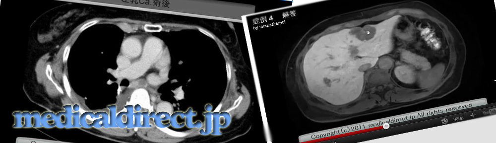
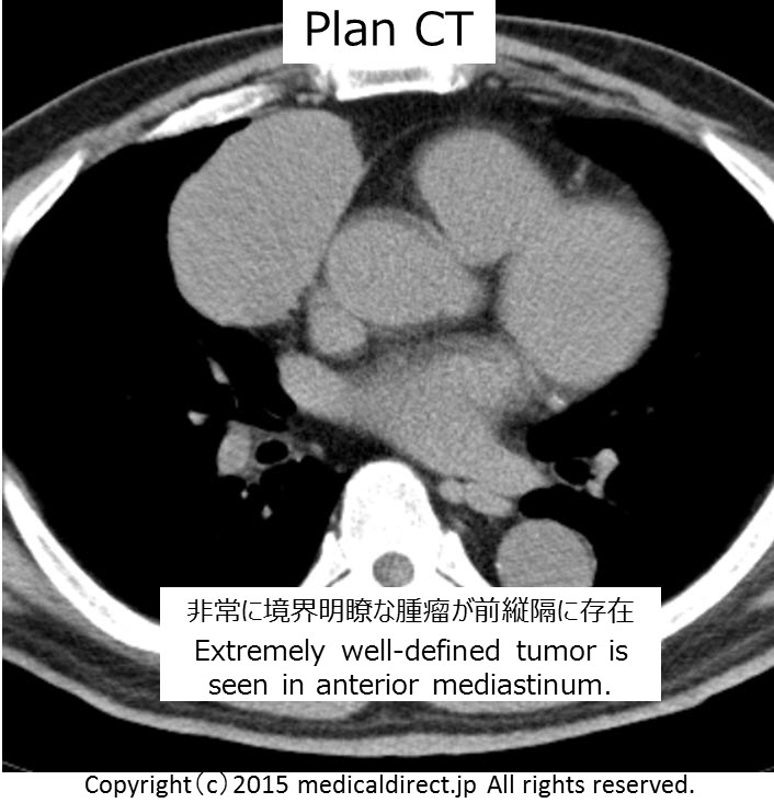
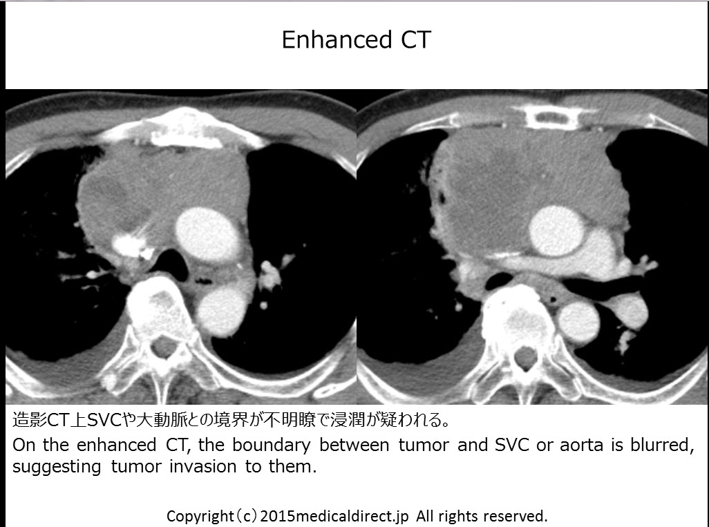
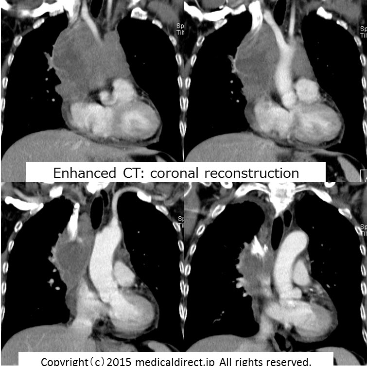
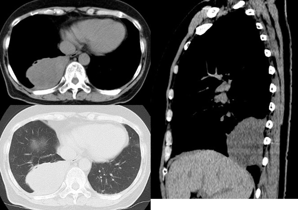
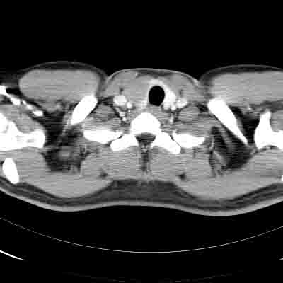
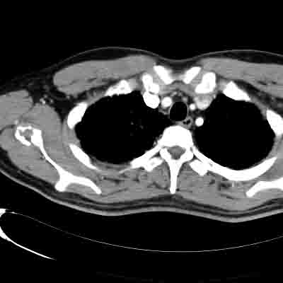
肺炎改善後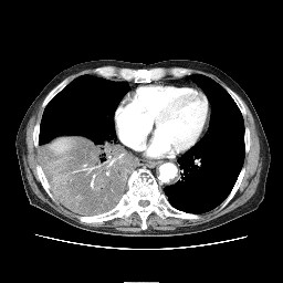 造影 CT 縦隔条件
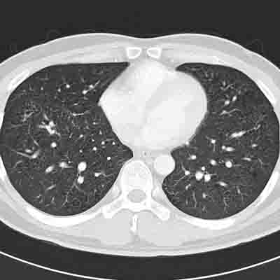
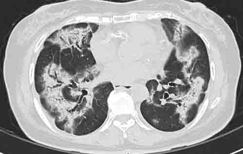
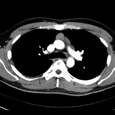
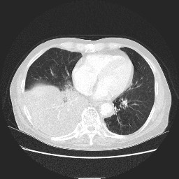 造影 CT 肺野条件
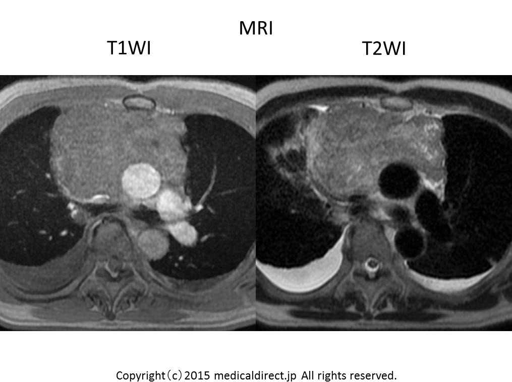
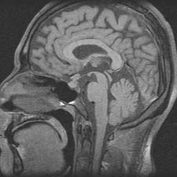 下垂体 MRI
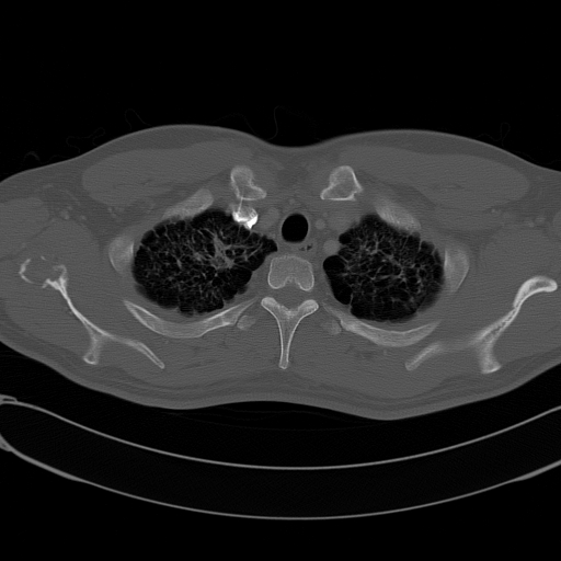
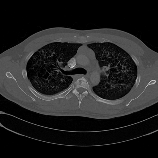
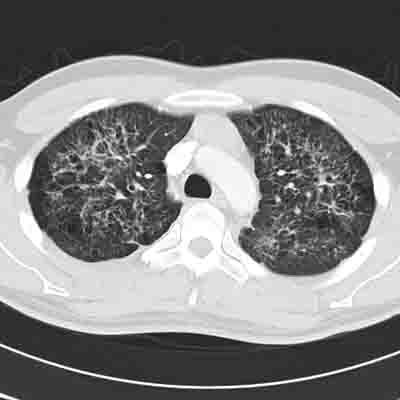
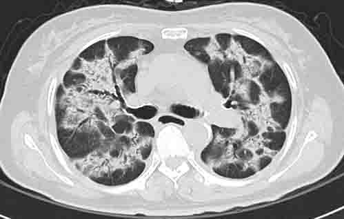
 肺野条件 2