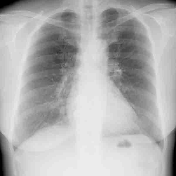
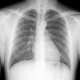
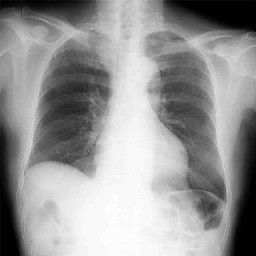
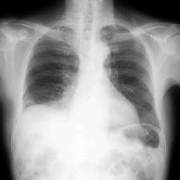
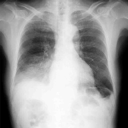
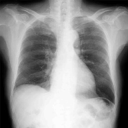
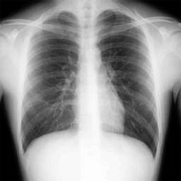
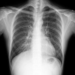
臨床症状:喘息の既往歴を有する10才代後半の男性 咳持続を主訴。
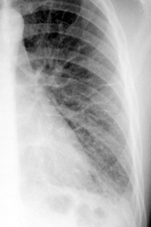
今回のエピソード胸部単純正面

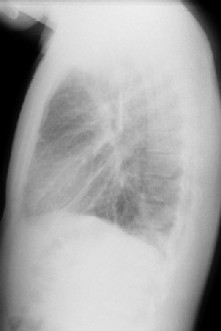
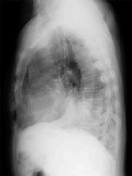
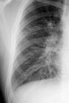
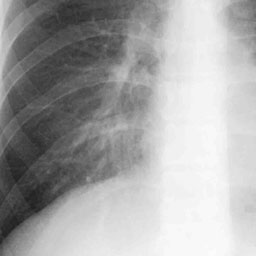
今回のエピソード胸部単純側面像
iphone movie
<Key words>
肺胞上皮癌、bronchioloalveolarCarcinoma、bronchoalveolar cell carcinoma、BAC、粘液産生、
CT angiogram sign,大葉性肺炎、大葉性陰影、陰影消長、悪性リンパ腫、malignant lymphoma
<文献リスト>
Trigaux JP, Gevenois PA, Goncette L, et al.
Bronchioloalveolar carcinoma: computed tomography findings.
Eur Respir J. 1996 Jan;9(1):11-6.
http://erj.ersjournals.com/cgi/reprint/9/1/11
Lee KS, Kim Y, Han J, et al.
Bronchioloalveolar carcinoma: clinical, histopathologic, and radiologic findings.
Radiographics. 1997 Nov-Dec;17(6):1345-57.
http://radiographics.rsna.org/content/17/6/1345.long
Aquino SL, Chiles C, Halford P.
Distinction of consolidative bronchioloalveolar carcinoma from pneumonia: do CT criteria work?
AJR Am J Roentgenol. 1998 Aug;171(2):359-63.
http://www.ajronline.org/cgi/reprint/171/2/359
Akira M, Atagi S, Kawahara M, Iuchi K, Johkoh T.
High-resolution CT findings of diffuse bronchioloalveolar carcinoma in 38 patients.
AJR Am J Roentgenol. 1999 Dec;173(6):1623-9.
http://www.ajronline.org/cgi/reprint/173/6/1623
Kim TH, Kim SJ, Ryu YH, et al.
Differential CT features of infectious pneumonia versus bronchioloalveolar carcinoma (BAC) mimicking pneumonia.
Eur Radiol. 2006 Aug;16(8):1763-8. Epub 2006 Jan 18.
Patsios D, Roberts HC, Paul NS, et al.
Pictorial review of the many faces of bronchioloalveolar cell carcinoma.
Br J Radiol. 2007 Dec;80(960):1015-23. Epub 2007 Oct 16.
http://bjr.birjournals.org/cgi/reprint/80/960/1015
Sawada E, Nambu A, Motosugi U, et al.
Localized mucinous bronchioloalveolar carcinoma of the lung: thin-section computed tomography and fluorodeoxyglucose positron emission tomography findings.
Jpn J Radiol. 2010 May;28(4):251-8. Epub 2010 May 29.
CT angiogram sign 関連
Im JG, Han MC, Yu EJ, et al.
Lobar bronchioloalveolar carcinoma: “angiogram sign” on CT scans.
Radiology. 1990 Sep;176(3):749-53.
Vincent JM, Ng YY, Norton AJ, Armstrong P.
CT “angiogram sign” in primary pulmonary lymphoma.
J Comput Assist Tomogr. 1992 Sep-Oct;16(5):829-31.
Murayama S, Onitsuka H, Murakami J, et al.
“CT angiogram sign” in obstructive pneumonitis and pneumonia.
J Comput Assist Tomogr. 1993 Jul-Aug;17(4):609-12.
Shah RM, Friedman AC.
CT angiogram sign: incidence and significance in lobar consolidations evaluated by contrast-enhanced CT.
AJR Am J Roentgenol. 1998 Mar;170(3):719-21.
http://www.ajronline.org/cgi/reprint/170/3/719
Maldonado RL.
The CT angiogram sign.
Radiology. 1999 Feb;210(2):323-4.
リンパ腫
Ooi GC, Chim CS, Lie AK, Tsang KW
Computed tomography features of primary pulmonary non-Hodgkin’s lymphoma.
Clin Radiol. 1999 Jul;54(7):438-43.
King LJ, Padley SP, Wotherspoon AC, Nicholson AG.
Pulmonary MALT lymphoma: imaging findings in 24 cases.
Eur Radiol. 2000;10(12):1932-8.
Lee DK, Im JG, Lee KS, Lee JS, Seo JB, Goo JM, Kim TS, Lee JW.
B-cell lymphoma of bronchus-associated lymphoid tissue (BALT): CT features in 10 patients.
J Comput Assist Tomogr. 2000 Jan-Feb;24(1):30-4.
<キー画像>
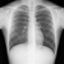
 1 年前正常時胸部単純写真 1 年前正常時胸部単純写真 |
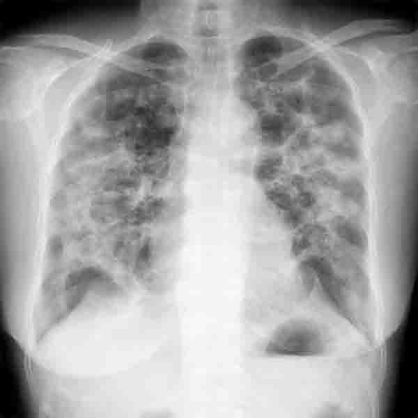
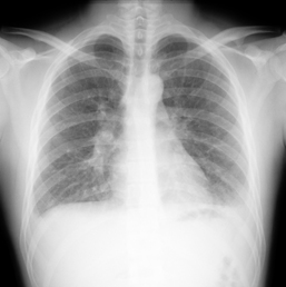
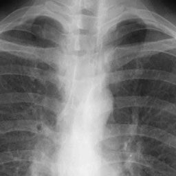
 10 ヶ月前肺炎発症時 10 ヶ月前肺炎発症時 |
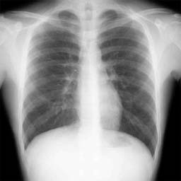
 今回の胸部単純写真正面像 今回の胸部単純写真正面像 |
 今回の胸部単純写真側面像 今回の胸部単純写真側面像 |
 肺炎改善後 肺炎改善後 |
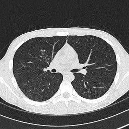
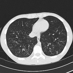
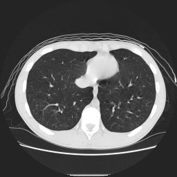
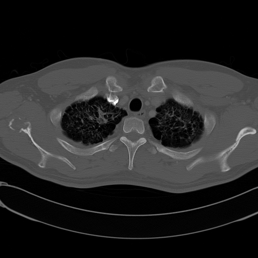
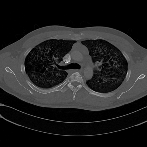
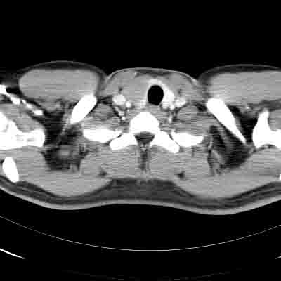
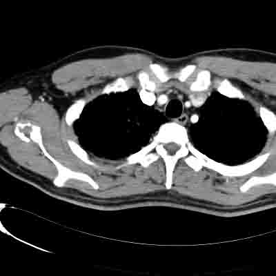
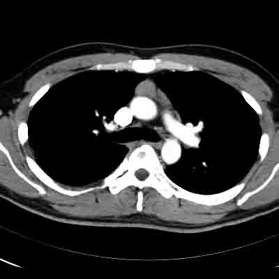
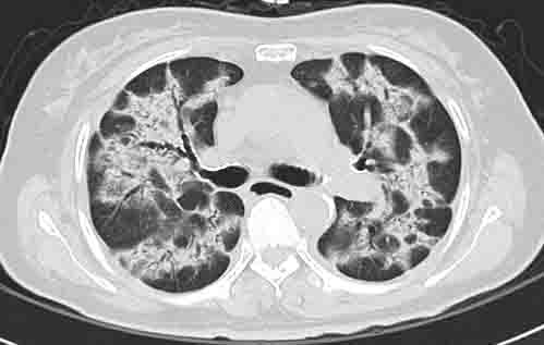
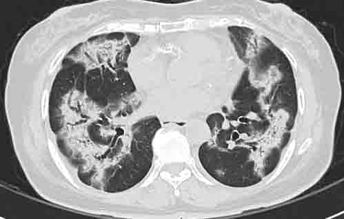
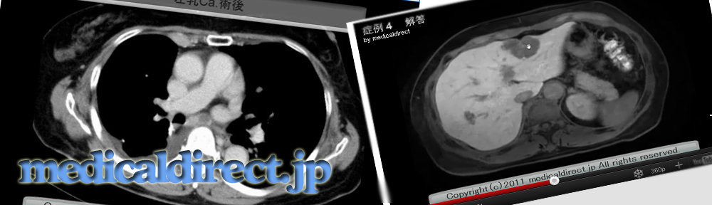
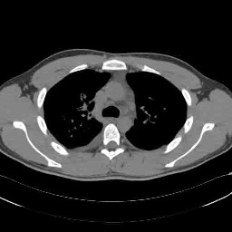
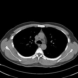
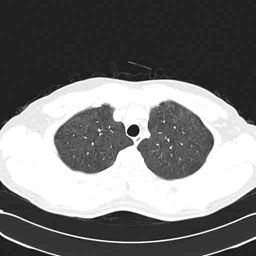
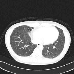
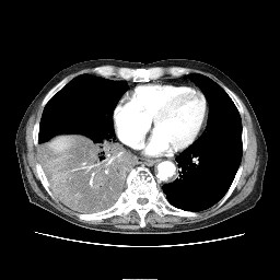 造影 CT 縦隔条件 造影 CT 縦隔条件 |
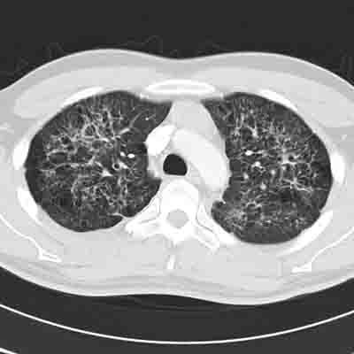
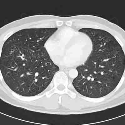
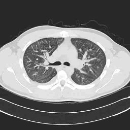
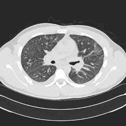
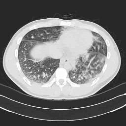
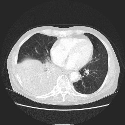 造影 CT 肺野条件 造影 CT 肺野条件 |
iphone movie
<key words>
びまん性汎細気管支炎、diffuse panbronchiolitis, DPB, 小葉中心性、分枝状 影、Y字、V字、細気管支炎, adult T-cell leukemia, lymphoma、非定型抗酸菌症、びまん性誤嚥性細気管支炎
<参考文献>
・DPBの論文
Akira M, Kitatani F, Lee YS, et al.
Diffuse panbronchiolitis: evaluation with high-resolution CT.
Radiology 1988 ;168:433-488.
Nishimura K, Kitaichi M, Izumi T, et al.
Diffuse panbronchiolitis: correlation of high-resolution CT and pathologic findings.
Radiology. 1992;184:779-785.
Akira M, Higashihara T, Sakatani M, et al.
Diffuse panbronchiolitis: follow-up CT examination.
Radiology. 1993 ;189:559-62.
Hansell DM.
Small airways diseases: detection and insights with computed tomography.
Eur Respir J. 2001 ;17:1294-313.
Xie GS, Li LY, Liu HR, et al.
Diffuse panbronchiolitis with histopathological confirmation among Chinese.
Chin Med J (Engl). 2004 ;117:1299-303.
・ATL関連の論文
Okada F, Ando Y, Kondo Y, et al.
Thoracic CT findings of adult T-cell leukemia or lymphoma.
AJR Am J Roentgenol. 2004 ;182:761-767.
Okada F, Ando Y, Yoshitake S, et al.
Pulmonary CT findings in 320 carriers of human T-lymphotropic virus type 1.
Radiology. 2006 Aug;240(2):559-64
<キー画像>
胸部単純半年前正面
|
今回のエピソードの 3 ヶ月後胸部単純正面 |
今回のエピソード胸部単純正面 |
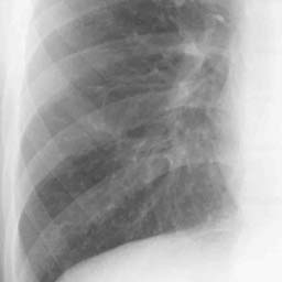
今回のエピソード トラムライン 拡大
|
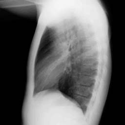
今回のエピソード胸部単純側面像
|
CT1 |
CT2
|
CT3 |
|
CT4(一ヵ月後のCT) |
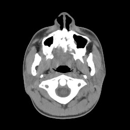
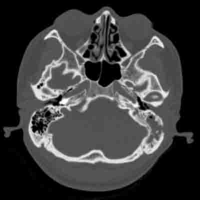
副鼻腔CT |
iphone用動画
<key word>喫煙、尿崩症、両肺対称性網状陰影、上肺優位の縮み、多発空洞性陰影、多発結節影、骨病変、Langerhans cell histiocytosis, LCH, eosinophilic granuloma, 好酸球性肉芽腫
<key画像>
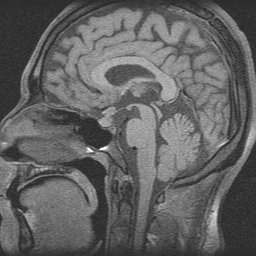 下垂体 MRI 下垂体 MRI |
骨病変 1 |
|
骨病変 2 |
縦隔条件 1 |
|
縦隔条件 2 |
縦隔条件 3 |
|
側頭骨 1 |
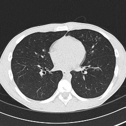
肺野条件 1 |
 肺野条件 2 肺野条件 2 |
肺野条件 3 |
<文献>
LCH肺
Abbott GF, Rosado-de-Christenson ML, et al.
From the archives of the AFIP: pulmonary Langerhans cell
histiocytosis.
Radiographics. 2004 May-Jun;24(3):821-41.
http://radiographics.rsna.org/content/24/3/821.full.pdf+html
Leatherwood DL, Heitkamp DE, Emerson RE.
Best cases from the AFIP: Pulmonary Langerhans cell
histiocytosis.
Radiographics. 2007 Jan-Feb;27(1):265-8.
http://radiographics.rsna.org/content/27/1/265.full.pdf+html
Sundar KM, Gosselin MV, Chung HL, Cahill BC.
Pulmonary Langerhans cell histiocytosis: emerging concepts
in pathobiology, radiology, and clinical evolution of
disease.
Chest. 2003 May;123(5):1673-83.
http://chestjournal.chestpubs.org/content/123/5/1673.full.pdf+html
Kulwiec EL, Lynch DA, Aguayo SM, Schwarz MI, King TE Jr.
Imaging of pulmonary histiocytosis X.
Radiographics. 1992 May;12(3):515-26.
http://radiographics.rsna.org/content/12/3/515.long
LCH縦隔
Donnelly LF, Frush DP.
Langerhans’ cell histiocytosis showing low-attenuation
mediastinal mass and cystic lung disease.
AJR Am J Roentgenol. 2000 Mar;174(3):877-8.
http://www.ajronline.org/cgi/content/full/174/3/877-a
LCH 全般
Meyer JS, Harty MP, Mahboubi S, Heyman S, Zimmerman RA,
Womer RB, Dormans JP, D’Angio GJ.
Langerhans cell histiocytosis: presentation and evolution
of radiologic findings with clinical correlation.
Radiographics. 1995 Sep;15(5):1135-46.
http://radiographics.rsna.org/content/15/5/1135.long
LCH 頭部
D’Ambrosio N, Soohoo S, Warshall C, Johnson A, Karimi S.
Craniofacial and intracranial manifestations of langerhans
cell histiocytosis: report of findings in 100 patients.
AJR Am J Roentgenol. 2008 Aug;191(2):589-97.
http://www.ajronline.org/cgi/reprint/191/2/589
Grois N, Prayer D, Prosch H, Lassmann H; CNS LCH
Co-operative Group.
Neuropathology of CNS disease in Langerhans cell
histiocytosis.
Brain. 2005 Apr;128(Pt 4):829-38. Epub 2005 Feb 10.
http://brain.oxfordjournals.org/cgi/reprint/128/4/829
Prayer D, Grois N, Prosch H, Gadner H, Barkovich AJ.
MR imaging presentation of intracranial disease associated
with Langerhans cell histiocytosis.
AJNR Am J Neuroradiol. 2004 May;25(5):880-91.
http://www.ajnr.org/cgi/reprint/25/5/880
LCH骨
Stull MA, Kransdorf MJ, Devaney KO.
Langerhans cell histiocytosis of bone.
Radiographics. 1992 Jul;12(4):801-23.
http://radiographics.rsna.org/content/12/4/801.long
Sartoris DJ, Parker BR.
Histiocytosis X: rate and pattern of resolution of osseous
lesions.
Radiology. 1984 Sep;152(3):679-84.
http://radiology.rsna.org/content/152/3/679.long
LCH甲状腺
Uchiyama M, Watanabe R, Ito I, Ikeda T.
Thyroid involvement in pulmonary langerhans cell
histiocytosis.
Intern Med. 2009;48(23):2047-8. Epub 2009 Dec 1.
http://www.jstage.jst.go.jp/article/internalmedicine/48/23/2047/_pdf
LCH肝
Radin DR.
Langerhans cell histiocytosis of the liver: imaging
findings.
AJR Am J Roentgenol. 1992 Jul;159(1):63-4.
<キーワード>
びまん性肺疾患、HRCT、volume loss, 容量減少、左右対称性陰影、bronchovascular bandle、気管支血管束、薬剤性肺障害、好酸球性肺炎、薬剤性好酸球性肺炎,traction bronchiectasis, 牽引性気管支拡張
<key画像>
|
2005 年 3 月正常時の単純写真 |
呼吸困難時、 2005 年末の胸部単純写真 |
|
呼吸困難時の胸部 CT1 |
呼吸困難時の胸部 CT2 |
| <参考文献>Souza CA, et al. Drug-induced eosinophilic pneumonia: high-resolution CT findings in 14 patients.AJR. 2006 ;186:368-73. http://www.ajronline.org/cgi/reprint/186/2/368Akira M, et al. Drug-induced pneumonitis: thin-section CT finding in 60 patients. Radiology. 2002;224 :852-860 http://radiology.rsna.org/content/224/3/852.full.pdf+htmlEllis, SJ, et al. Drug-induced lung disease: high-resolution CT findings. AJR. 2000;175 :1019-1024 http://www.ajronline.org/cgi/reprint/175/4/1019Cleverley JR, et al. Drug-induced lung disease: high-resolution CT and histological findings.Clin Radiol. 2002;57:292-9. Johkoh T, et al. Eosinophilic lung diseases: diagnostic accuracy of thin-section CT in 111 patients.Radiology. 2000;216 :773-80. http://radiology.rsna.org/content/216/3/773.full.pdf+html*パナルジンが原因の間質性肺炎の症例報告論文 Oon PC, et al. Diffuse alveolar damage associated with ticlopidine use: a case report.J Formos Med Assoc. 2003;102 :262-5.Nakamura R, et al. Interstitial pneumonia induced by ticlopidine.Circ J. 2002;66 :773-776. http://www.jstage.jst.go.jp/article/circj/66/8/773/_pdfWatanebe M, et al. Multiple pulmonary nodules due to ticlopidine-induced pneumonitis Nihon Kokyuki Gakkai Zasshi. 1999;37 :841-5Alonso-Martinez JL, et al. Bronchiolitis obliterans-organizing pneumonia caused by ticlopidine.Ann Intern Med. 1998;129 :71-2 http://www.annals.org/content/129/1/71.3.full.pdf+html |
急性好酸球性肺炎、acute eosinophilic pneumonia, eosinophilic lung diseases、カーリーライン、Kerley line, Kerley B line、広義間質、bronchovascular bundle, septal line, 小葉間隔壁、non cardiogenic edema、薬剤性肺障害
<key画像>
|
正常時 |
発症時 |
|
|
 |
 |
 |
 |
 |
 |
<参考文献>
急性好酸球性肺炎文献集
Jeong YJ, Kim KI, Seo IJ, Lee CH, Lee KN, Kim KN, Kim JS, Kwon WJ.
Eosinophilic lung diseases: a clinical, radiologic, and pathologic overview.
Radiographics. 2007 May-Jun;27(3):617-37; discussion 637-9.
http://radiographics.rsna.org/content/27/3/617.full.pdf+html
AEP症例報告 Abe K, Yanagi S, Imadsu Y, Sano A, Iiboshi H, Mukae H, Matsukura S.
Acute eosinophilic pneumonia with fine nodular shadows.
Intern Med. 2003 Jan;42(1):88-91.
Shiota Y, Kawai T, Matsumoto H, Hiyama J, Tokuda Y, Marukawa M, Ono T, Mashiba H.
Acute eosinophilic pneumonia following cigarette smoking.
Intern Med. 2000 Oct;39(10):830-3.
症例報告の直截リンクは張れませんが、Freeで取れます。画像は決してよいと言えません。
画像はpoorだが、臨床的なこと知りたい場合
Philit F, Etienne-Mastroianni B, Parrot A, Guerin C, Robert D, Cordier JF.
Idiopathic acute eosinophilic pneumonia: a study of 22 patients.
Am J Respir Crit Care Med. 2002 Nov 1;166(9):1235-9.
http://ajrccm.atsjournals.org/cgi/reprint/166/9/1235
参考として 薬剤性肺炎の論文
Rossi SE, Erasmus JJ, McAdams HP, Sporn TA, Goodman PC.
Pulmonary drug toxicity: radiologic and pathologic manifestations.
Radiographics. 2000 Sep-Oct;20(5):1245-59. Review.
http://radiographics.rsna.org/content/20/5/1245.full.pdf+html
iphone movie
key words
夏型過敏性肺臓炎、過敏性肺臓炎、hypersensitivity pneumonitis, summer-type hypersensitivity pneumonitis, びまん性スリガラス陰影、小葉中心性、粒状陰影、広範囲、スリガラス陰影、吸引抗原、ニューモシスチス肺炎、縦隔気腫
文献
過敏性肺臓炎
Silva CI, Churg A, Muller NL.
Hypersensitivity pneumonitis: spectrum of high-resolution CT and pathologic findings.
AJR Am J Roentgenol. 2007 ;188:334-44.
http://www.ajronline.org/cgi/reprint/188/2/334
Hansell DM, Wells AU, Padley SP, Muller NL.
Hypersensitivity pneumonitis: correlation of individual CT patterns with functional abnormalities.
Radiology. 1996; 199:123-128.
http://radiology.rsna.org/content/199/1/123.long
Glazer CS, Rose CS, Lynch DA.
Clinical and radiologic manifestations of hypersensitivity pneumonitis.
J Thorac Imaging. 2002 ;17:261-272.
Patel RA, Sellami D, Gotway MB, et al.
Hypersensitivity pneumonitis: patterns on high-resolution CT.
J Comput Assist Tomogr. 2000 ;24:965-970.
Lynch DA, Newell JD, Logan PM, et al.
Can CT distinguish hypersensitivity pneumonitis from idiopathic pulmonary fibrosis?
AJR Am J Roentgenol. 1995 ;165):807-811.
http://www.ajronline.org/cgi/reprint/165/4/807
Lynch DA, Rose CS, Way D, King TE Jr.
Hypersensitivity pneumonitis: sensitivity of high-resolution CT in a population-based study.
AJR Am J Roentgenol. 1992 ;159:469-472.
http://www.ajronline.org/cgi/reprint/159/3/469
Akira M, Kita N, Higashihara T, et al.
Summer-type hypersensitivity pneumonitis: comparison of high-resolution CT and plain radiographic findings.
AJR Am J Roentgenol. 1992 ;158:1223-8.
http://www.ajronline.org/cgi/reprint/158/6/1223
Silver SF, Muller NL, Miller RR, et al.
Hypersensitivity pneumonitis: evaluation with CT. Radiology 1989; 173:441-445
http://radiology.rsna.org/content/173/2/441.long
慢性過敏性肺臓炎
Silva CI, Muller NL, Lynch DA, et al.
Chronic hypersensitivity pneumonitis: differentiation from idiopathic pulmonary fibrosis and nonspecific interstitial pneumonia by using thin-section CT.
Radiology. 2008 ;246:288-297.
http://radiology.rsna.org/content/246/1/288.full.pdf+html
Sahin H, Brown KK, Curran-Everett D, et al.
Chronic hypersensitivity pneumonitis: CT features comparison with pathologic evidence of fibrosis and survival.
Radiology. 2007 ;244:591-598.
http://radiology.rsna.org/content/244/2/591.full.pdf+html
Adler BD,Padley SP,Muller NL,et al.
Chronic hypersensitivity pneumonitis: high-resolution CT and radiographic features in 16 patients.
Radiology. 1992 ;185:91-95.
http://radiology.rsna.org/content/185/1/91.long
Buschman DL,Gamsu G,Waldron JA Jr,et al.
Chronic hypersensitivity pneumonitis: use of CT in diagnosis.
AJR Am J Roentgenol. 1992 ;159:957-960.
http://www.ajronline.org/cgi/reprint/159/5/957
2次小葉についての論文
Webb WR.
Thin-section CT of the secondary pulmonary lobule: anatomy and the image–the 2004 Fleischner lecture.
Radiology. 2006 ;239:322-38. Epub 2006 Mar 16.
http://radiology.rsna.org/content/239/2/322.full.pdf+html
key画像
 |
 |
 |
 |
 |
 |
臨床症状:喘息の既往歴を有する10才代後半の男性 咳持続を主訴。
今回のエピソード胸部単純正面
今回のエピソード胸部単純側面像