62-year-old man with abnormal shadow in right lower lung field during follow up of brain infarction.

62才男性
脳梗塞フォロー中右下肺野に異常陰影指摘

まだ問題を見ていない先生はこちら

Case 1

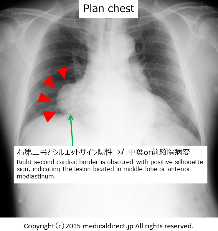
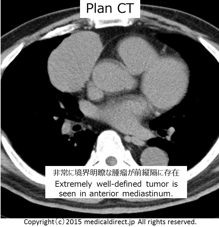
62-year-old man with abnormal shadow in right lower lung fieldduring follow up of brain infarction.

iPad version


You tube version


今回は解答編 バージョン1です。症例の解説が主体です。

解答編バージョン2で胸腺腫総まとめしています。

胸腺腫の総まとめビデオはこちら 10分余った時間で勉強してください。

Key words: Mediastinal tumor, Thymoma, thymoma, thymic tumor, 縦隔腫瘍、胸腺腫、胸腺腫瘍、WHO classification, WHO分類

Key images:

Case 1  62M Type A

Type A thymoma 62M Chest plain 1Type A thymoma 62M CT plain 1Type A thymoma 62M MRI 1Type A thymoma 62M MRI 2Type A thymoma 62M MRI 3Type A thymoma 62M MRI 4

Case 2  69MType B3 Thymoma 69M Chest plain1 Type B3 Thymoma 69M CT1Type B3 Thymoma 69M CT2Type B3 Thymoma 69M MRI1Type B3 Thymoma 69M MRI 2.6

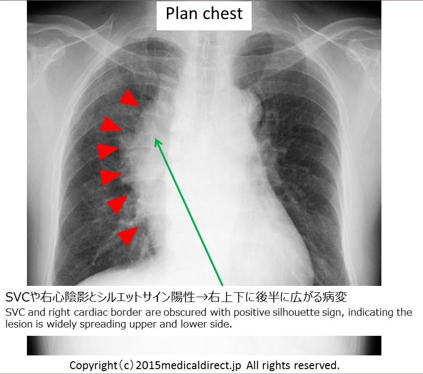
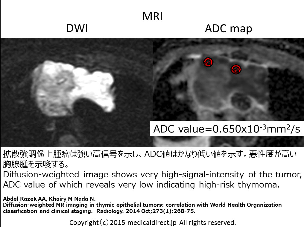
胸腺腫: ADCとWHO分類との対比論文はこちら

1.

Abdel RazekAA, KhairyM Nada N.

Diffusion-weighted MR imaging in thymic epithelial tumors: correlation with World Health Organization classification and clinical staging. Radiology. 2014 Oct;273(1):268-75.

2.

Priola AM, Priola SM.

Usefulness of Diffusion-weighted MR Imaging in Predicting Masaoka-Koga Clinical Staging of Thymic Epithelial Tumors by Using the Apparent Diffusion Coefficient.  Radiology. 2015 Mar;274(3):936-7.


コメントは受け付けていません。