回答は2週間以内に
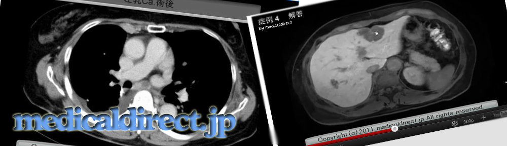
70歳代男性、管理職 喫煙20本/日 機会飲酒
返信
回答は2週間以内に
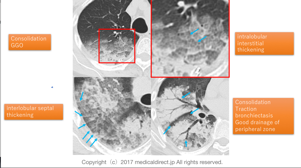
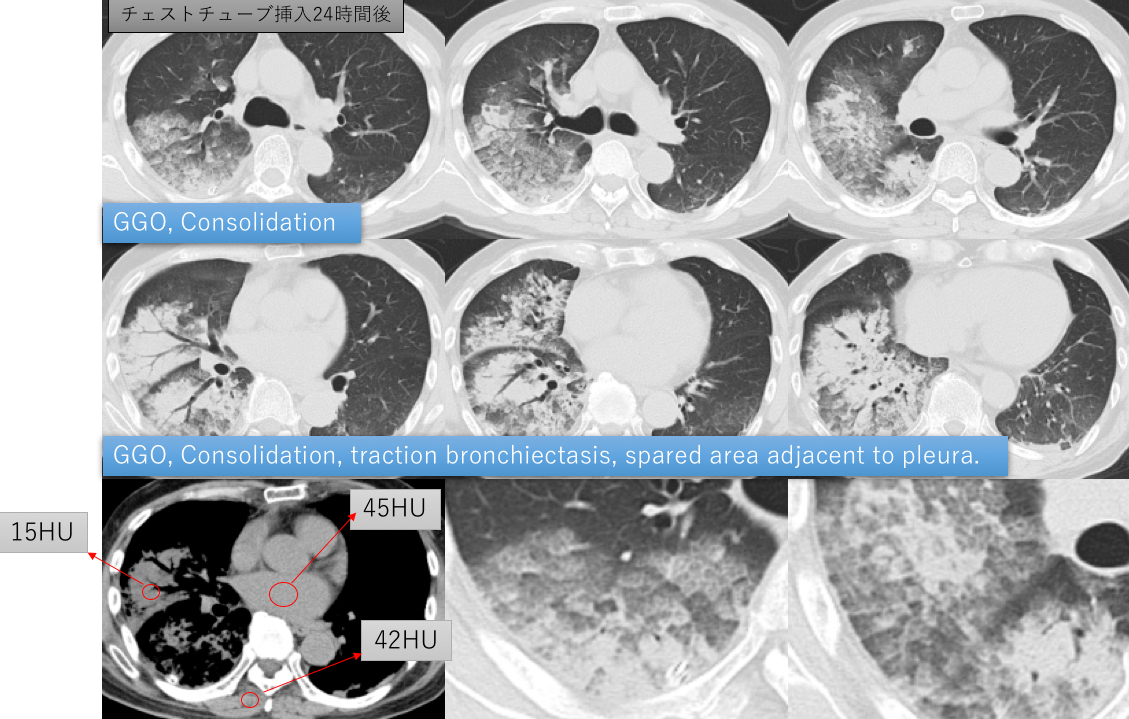
Key words: re-expansion pulmonary edema, REPE, GGO, consolidation,
intralobular interstitial thickening, interlobular septal thickening, infiltration 浸潤影
肺胞出血、肺炎、COP
Reference:
Jun Hyun Baik, et al.
High-Resolution CT Findings of Re-Expansion Pulmonary Edema
Korean J Radiol 2010;11:164-168
Key images
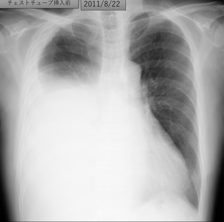
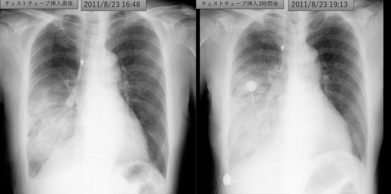

右胸水大量貯留に対するドレナージを施行すると・・・
果たして何が生じたのか?
また、左肺の変化は説明できるのか?
がこの問題のポイントです。
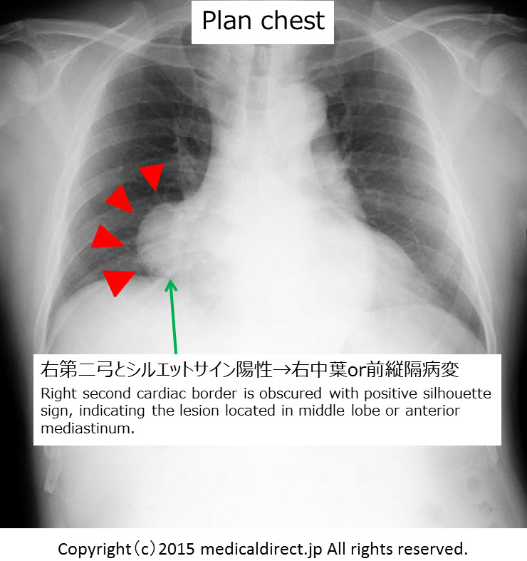
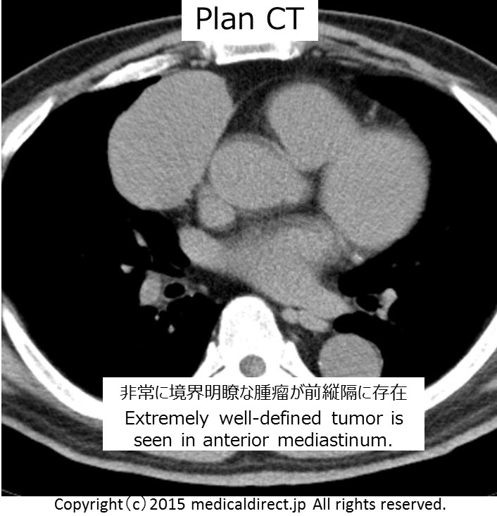
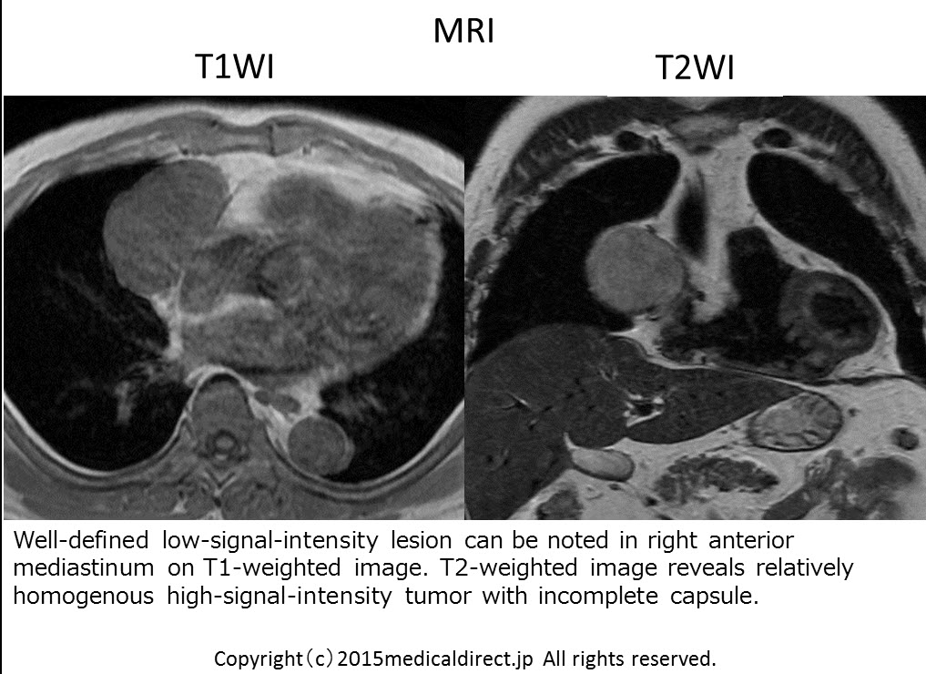
What is your diagnosis?
Why the consolidation can be appeared?
回答はこちら
Keywords: ABPA, Allergic bronchopulmonary aspergillosis, アレルギー性気管支肺アスペルギルス症, 喘息、asthma, mucoid impaction, Bronchiectasis, Mucoceles, High attenuation mucus
イチロウのみのオリジナル最新文献レビューを含めた動画はもう少しお待ちください。
注)エキスパートシリーズの肺は残念ながら途中で終わっています。
エキスパートがそれ以上のホームページアップを望まなかったからです。
なので私がそのとき習ったこととさらに文献をなどを引用し肉付けして
代わりに動画でアップしていきます。お楽しみに。
Thymoma teaching, 胸腺腫 まとめ
*動画の題名が縦隔腫瘍になっていますが、実際は胸腺腫のまとめです。
iPad version
You tube version
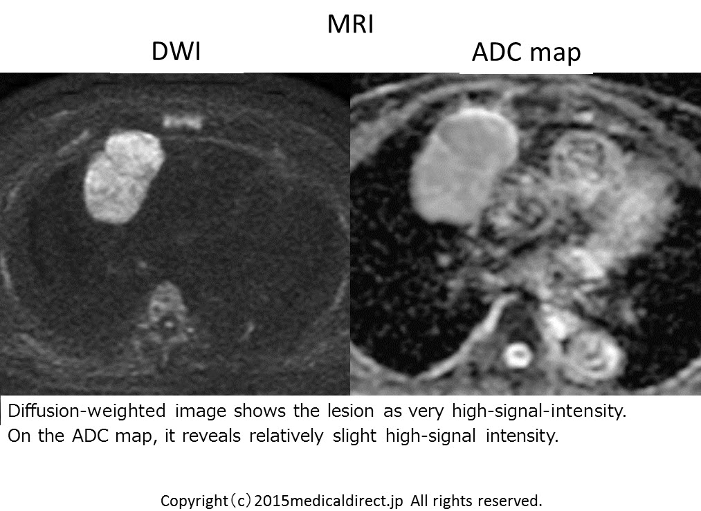
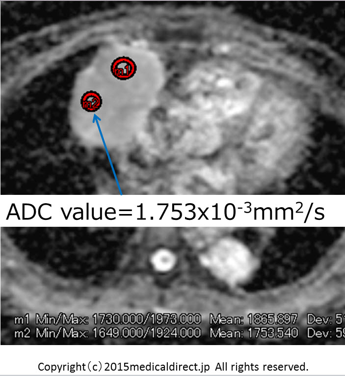
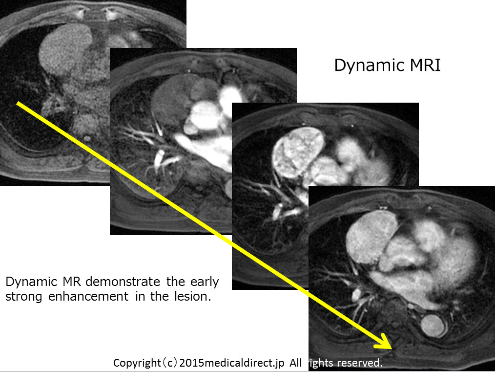
Key words: Thymoma, 胸腺腫、anterior mediastinal tumor, 前縦隔腫瘍, WHO classification, WHO分類、Masaoka classification, 正岡分類、胸腺癌, thymic cancer, Diffusion weighted image, DWI, 拡散強調画像
まとめ:黒色は非浸潤性胸腺腫、赤色はより浸潤性、紫色はより癌を示唆
References:
1. Priola AM, Priola SM, Di Franco M, et al.Computed tomography and thymoma: distinctive findings in invasive and noninvasive thymoma and predictive features of recurrence.
RadiolMed. 2010 Feb;115(1):1-21.
2. Tomiyama N, Müller NL, Ellis SJ, et al.
Invasive and noninvasive thymoma: distinctive CT features.
J Comput Assist Tomogr. 2001 May-Jun;25(3):388-93.
3. Jeong YJ, Lee KS, Kim J, et al.
Does CT of thymic epithelial tumors enable us to differentiate histologic subtypes and predict prognosis?
Am J Roentgenol. 2004 Aug;183(2):283-9.
4. Harris K, Elsayegh D, Azab B, et al.10.1186/1477-7819-9-95.
Thymoma calcification: is it clinically meaningful?
World J SurgOncol. 2011 Aug 23;9:95.
5. Hu YC, Wu L, Yan LF, et al.
Predicting subtypes of thymic epithelial tumors using CT: new perspective based on a comprehensive analysis of 216 patients.
Sci Rep. 2014 Nov 10;4:6984.
6. Sadohara J, Fujimoto K, Müller NL, et al.
Thymic epithelial tumors: comparison of CT and MR imaging findings of low-risk thymomas, high-risk thymomas, and thymic carcinomas.
Eur J Radiol. 2006 Oct;60(1):70-9.
7. Inoue A, Tomiyama N, Fujimoto K, et al.
MR imaging of thymic epithelial tumors: correlation with World Health Organization classification.
Radiat Med.2006 Apr;24(3):171-81.
8. Han J, Lee KS, Yi CA, et al.
Thymic epithelial tumors classified according to a newly established WHO scheme: CT and MR findings.
Korean J Radiol. 2003 Jan-Mar;4(1):46-53.
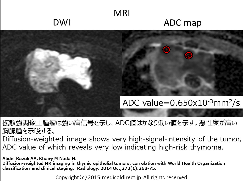
★Diffusion-weighted images in thymomas.
1. Abdel Razek AA, Khairy M Nada N.
Diffusion-weighted MR imaging in thymic epithelial tumors: correlation with World Health Organization classification and clinical staging.
Radiology. 2014 Oct;273(1):268-75.
2. Priola AM, Priola SM, Giraudo MT, et al.
Chemical-shift and diffusion-weighted magnetic resonance imaging of thymus in myasthenia gravis: usefulness of quantitative assessment.
Invest Radiol. 2015 Apr;50(4):228-38
3. Seki S, Koyama H, Ohno Y, et al.
Diffusion-weighted MR imaging vs. multi-detector row CT: Direct comparison of capability for assessment of management needs for anterior mediastinal solitary tumors
Eur J Radiol. 2014 May;83(5):835-42.
4. Shin KE, Yi CA, Kim TS, et al.
Diffusion-weighted MRI for distinguishing non-neoplastic cysts from solid masses in the mediastinum: problem-solving in mediastinal masses of indeterminate internal characteristics on CT. EurRadiol. 2014 Mar;24(3):677-84.
EurRadiol. 2014 Mar;24(3):677-84.
62才男性
脳梗塞フォロー中右下肺野に異常陰影指摘
Case 1
62-year-old man with abnormal shadow in right lower lung fieldduring follow up of brain infarction.
iPad version
You tube version
今回は解答編 バージョン1です。症例の解説が主体です。
胸腺腫の総まとめビデオはこちら 10分余った時間で勉強してください。
Key words: Mediastinal tumor, Thymoma, thymoma, thymic tumor, 縦隔腫瘍、胸腺腫、胸腺腫瘍、WHO classification, WHO分類
Key images:
Case 1 62M Type A
Case 2 69M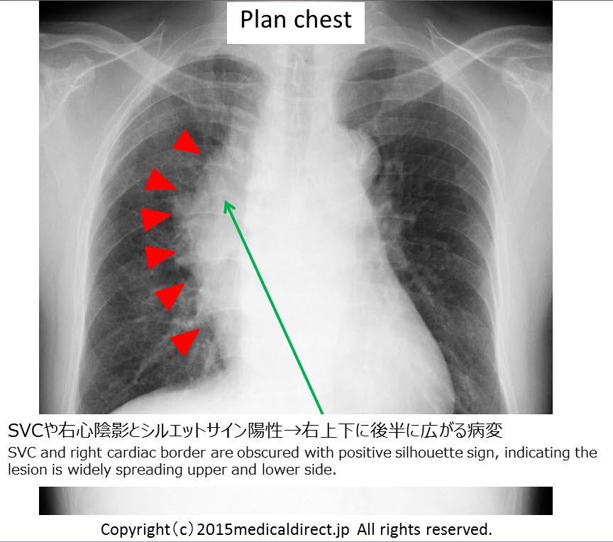
胸腺腫: ADCとWHO分類との対比論文はこちら
1.
Abdel RazekAA, KhairyM Nada N.
2.