Thymoma teaching, 胸腺腫 まとめ
*動画の題名が縦隔腫瘍になっていますが、実際は胸腺腫のまとめです。
iPad version
You tube version
Key words: Thymoma, 胸腺腫、anterior mediastinal tumor, 前縦隔腫瘍, WHO classification, WHO分類、Masaoka classification, 正岡分類、胸腺癌, thymic cancer, Diffusion weighted image, DWI, 拡散強調画像
まとめ:黒色は非浸潤性胸腺腫、赤色はより浸潤性、紫色はより癌を示唆
- 大きい
- 被膜(より完全)、隔壁
- 境界明瞭、不明瞭
- 分葉状、不整
- 出血、壊死、嚢胞性変化
- 石灰化
- 不均一造影効果
- 造影効果が高いか否か
- T2WI上不均一
- T2WI上腫瘍内の低信号
- 胸膜播種
- 縦隔脂肪浸潤
- 大血管浸潤
- LN腫大
- 線維性隔壁:AB、B
- MRIは被膜、隔壁、出血の検出でCTより優れる
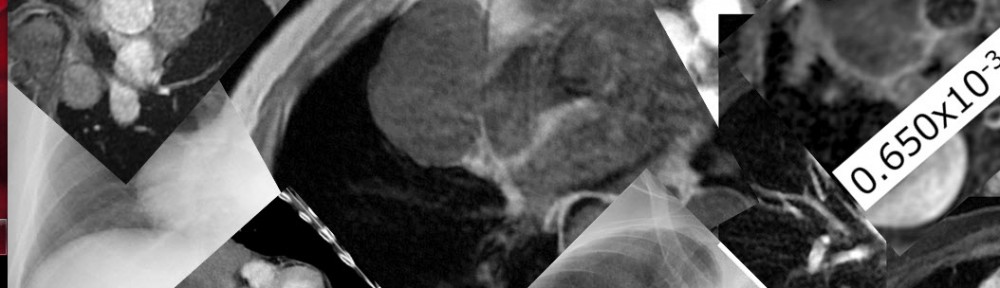
- ADC値はWHO分類、正岡分類と相関:ADC値低いとより悪性度高い
References:
1. Priola AM, Priola SM, Di Franco M, et al.Computed tomography and thymoma: distinctive findings in invasive and noninvasive thymoma and predictive features of recurrence.
RadiolMed. 2010 Feb;115(1):1-21.
2. Tomiyama N, Müller NL, Ellis SJ, et al.
Invasive and noninvasive thymoma: distinctive CT features.
J Comput Assist Tomogr. 2001 May-Jun;25(3):388-93.
3. Jeong YJ, Lee KS, Kim J, et al.
Does CT of thymic epithelial tumors enable us to differentiate histologic subtypes and predict prognosis?
Am J Roentgenol. 2004 Aug;183(2):283-9.
4. Harris K, Elsayegh D, Azab B, et al.10.1186/1477-7819-9-95.
Thymoma calcification: is it clinically meaningful?
World J SurgOncol. 2011 Aug 23;9:95.
5. Hu YC, Wu L, Yan LF, et al.
Predicting subtypes of thymic epithelial tumors using CT: new perspective based on a comprehensive analysis of 216 patients.
Sci Rep. 2014 Nov 10;4:6984.
6. Sadohara J, Fujimoto K, Müller NL, et al.
Thymic epithelial tumors: comparison of CT and MR imaging findings of low-risk thymomas, high-risk thymomas, and thymic carcinomas.
Eur J Radiol. 2006 Oct;60(1):70-9.
7. Inoue A, Tomiyama N, Fujimoto K, et al.
MR imaging of thymic epithelial tumors: correlation with World Health Organization classification.
Radiat Med.2006 Apr;24(3):171-81.
8. Han J, Lee KS, Yi CA, et al.
Thymic epithelial tumors classified according to a newly established WHO scheme: CT and MR findings.
Korean J Radiol. 2003 Jan-Mar;4(1):46-53.
★Diffusion-weighted images in thymomas.
1. Abdel Razek AA, Khairy M Nada N.
Diffusion-weighted MR imaging in thymic epithelial tumors: correlation with World Health Organization classification and clinical staging.
Radiology. 2014 Oct;273(1):268-75.
2. Priola AM, Priola SM, Giraudo MT, et al.
Chemical-shift and diffusion-weighted magnetic resonance imaging of thymus in myasthenia gravis: usefulness of quantitative assessment.
Invest Radiol. 2015 Apr;50(4):228-38
3. Seki S, Koyama H, Ohno Y, et al.
Diffusion-weighted MR imaging vs. multi-detector row CT: Direct comparison of capability for assessment of management needs for anterior mediastinal solitary tumors
Eur J Radiol. 2014 May;83(5):835-42.
4. Shin KE, Yi CA, Kim TS, et al.
Diffusion-weighted MRI for distinguishing non-neoplastic cysts from solid masses in the mediastinum: problem-solving in mediastinal masses of indeterminate internal characteristics on CT. EurRadiol. 2014 Mar;24(3):677-84.
EurRadiol. 2014 Mar;24(3):677-84.