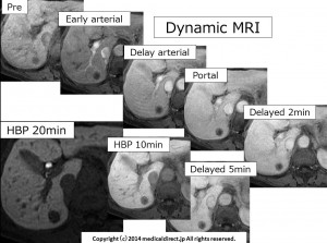
EOBプリモビスト症例集9例目解答編

70才台女性、便秘症、肝内SOL指摘

問題を見ていない先生はこちら

この症例を10分間聞くことで先生が得られる結果

①いつも苦労していた血管腫との鑑別がある方法によって簡単になります。

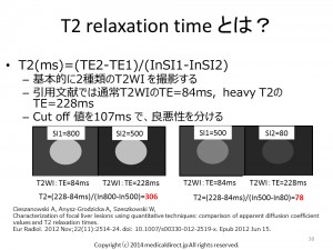
 実はこの方法は昔から使われていましたが、最近のある論文で再び見直されました。

CieszanowskiA, Anysz-Grodzicka A, Szeszkowski W,

Characterization of focal liver lesions using quantitative techniques: comparison of apparent diffusion coefficient values and T2 relaxation times.

EurRadiol. 2012 Nov;22(11):2514-24. doi: 10.1007/s00330-012-2519-x. Epub 2012 Jun 15.

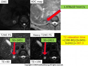
この論文では ADC map(accuracy 81%) より T2 relaxation time (同 89%)の方が使えると言っています。

論文は2012年とこのVideo をアップしたときには非常に新しいものです。

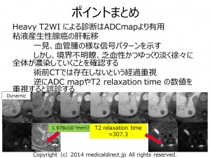
②上記を知ったうえで、今回の症例のように注意が非常に必要な症例 つまり例外を知ることも重要です。日常診療で輝きを増すには例外を明確に知っているかで決まります。T2 relaxation time を使いこなし、例外も知ることでEOB-MRIでの血管腫との鑑別が容易になります。

③粘液癌、粘液産生性腺癌 vs. 血管腫は 永遠のテーマであるが、実は症例をじっくり眺めているとヒントが得られる可能性もあります。粘液癌、粘液産生性腺癌はT2 relaxation time を使わなくても形態的にも違いが分かります(文献はまだありませんが)

さあ、たった10分です。余った時間で聞いてみてください。

ちなみにこの問題は、ある放射線医学教室の後期研修医に

問題としてだして いくつかある問題のうち9%しか正解の得られなかった難問です。

iPhone, iPad の先生はこちら

You tube version はこちら


Key words: 粘液産生性腺癌、Mucinous adenocarcinoma, mucinous cyst adenocarcinoma, 肝転移、liver metastases, 血管腫、hemangioma, T2 relaxation time, ADC map

Key images.

HP 添付したkey images 1HP 添付したkey images 2HP 添付したkey images 3HP 添付したkey images 4Similar cases  類似症例: 1週間以内にスライドアップする予定です。


EOBプリモビスト第8回 回答

60才台男性
糖尿病、慢性肝炎 治療中
S6に超音波上SOLを指摘される

まず、問題を見てからという先生はこちら

この10分の1症例の回答ビデオを見ることであなたが得られるベネフィット

①先生の意識次第では単純CTが意外に役に立つことを知れます。これにより今後決して単純CTを撮影から外してしまったり、読影に使わなかったりはしないでしょう。

②inphase, opposed phase をよく見ることにより、病変内部の性状を詳細に見れるようになるでしょう。より病理に近い診断を頭の中に描くことが可能となります。

③Dynamic MRIをよくよくみると、病理分化度の違いに気が付かれることでしょう。その瞬間もし先生が、「おや?」 と思われたなら、かなり肝臓画像診断に精通している先生です。「俺はわかったからもうビデオは見ない?」 いえ、ちょっとその先の3分間も見てみてください。

④肝細胞相で結節全体の大きさがわかるので、opposed phase と対比してみると結節が2種類の病理像から構成されていることがわかります。この対比はHCC診断とくに早期高分化型あるいは早期から多血化に脱分化する状況において重要です。

⑤HCCの多段階発癌という現象が実際の画像でどのような状況というか所見を作り出すことになるのかを解説しています。これを見ることで先生は実は今回の症例が変わった症例なんだと言う事を認識できます。ビデオ6:25 あたり。ではいったい通常のパターンとはどういう画像所見になるのでしょうか? ビデオ8:10 あたり

iPad version

You tube version

key words: 早期高分化型肝癌、早期高分化型HCC、脂肪化、肝癌の 多段階発育、early hepatocellular carcinoma, early hepatoma, fatty change, fatty metamorphosis, multistep carcinogenesis

問題はこちら

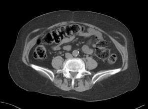
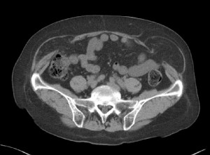
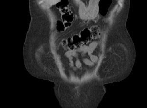
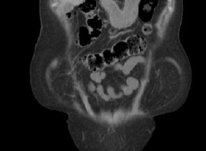
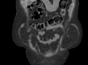
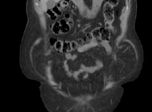
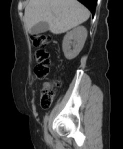
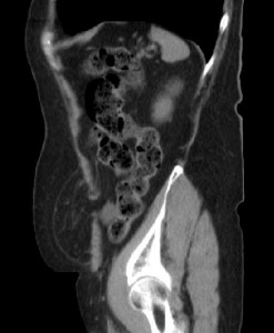
Abdominal wall hernia Answer 解答編

Abdominal wall hernia: Spiegelian hernia  半月状線ヘルニア

問題をみていないので、問題を見てからこの回答を見る

iPad version


You tube version

key words: Abdominal wall hernia, 腹壁ヘルニア、Spiegelian hernia, 半月状線ヘルニア、hernia, ヘルニア、surgery, 手術

Spiegelian hernia まとめ

  • 半月状線ヘルニア:腹横筋線維が腱膜に移行する腹直筋外縁の半月状線,すなわちSpigel腱膜に発生する稀なヘルニア
  • 半月状線の脆弱性、上前腸骨棘を結ぶ線から6cm上方までに発生しやすい=SH belt
  • 平均年齢60才台、女性優位
  • 左優位、両側性は稀
  • 修復術を行った腹壁ヘルニア中の頻度は1%未満
  • 原因として外科手術や極度の肥満など
  • 外腹斜筋腱膜をこえるか否かで2つに分類

–Interstitial SH: closed loop Small bowel obstruction なり易い

–Subcutaneous SH

  • CTで100%の感度とPPV.

Key images

半月状線とはどこ?1半月状線とはどこ?2

CT0050CT0056

CT0009CT0010CT0011CT0012CT0015CT0048文献:

1.Martine M, Paquette B, Badet N, et al. Spiegelian hernia: CT findings and clinical relevance. Abdom imaging. 2013; 38: 260-264

2. Moles Morenilla L, DocoboDurántez F, Mena Robles J,etal. Spiegelianhernia in Spain. An analysis of 162 cases. Rev EspEnferm Dig. 2005 May;97(5):338-47.

3. Light D, Chattopadhyay D, Bawa S. Radiological and clinical examination in the diagnosis of Spiegelian hernia. Ann R CollSurgEngl 2013; 95:98-10

4. Dabbas N, Adams K, Pearson K, et al.  Frequency of abdominal wall hernias: is classical teaching out of date?   JRSM Short Rep.2011 Jan 19;2(1):5. doi: 10.1258/shorts.2010.010071.

5.  塩田喜代美、植木孝宣、青井重善、他。CTにて術前診断した半月状線ヘルニアの1例  日臨外会誌 . 2002: 63: 1308-1311

English summary

Summary of Spiegelian hernia

  • Spigelian hernias (SH) are rarely reported abdominal wallhernias, which arise from the Spigelianfascia. It lies along the semilunar line lateral to the rectus abdominis muscle.
  • SH is due to a weakness of the spiegelian fascia.
  • Most of the SH cases occur within the zone, so-called SH belt, which is a transverse band between the line joining both anterior superior iliac spines and parallel line lying 6-cm cranial to it.
  • Mean age is 60-70 year-old. Female domination.
  • Left side is dominant. Bilateral SH is very rare (2%).
  • The incidence of SH among abdominal wall hernia repair surgery is less than 1%.
  • Major acquired risk factors of SH are a history of abdominal surgery and obesity.
  • Two types of SH are interstitial SH and subcutaneous SH. Closed loop small bowel obstruction is statistically associated with interstitial SH.
  • CT has a 100% sensitivity and 100% PPV todiagnose SH.


27-year-old man with knee injury

回答

Meniscofemoral ligament (MFL)

iPad version.


You tube version.


Key words: 膝、膝関節、正常構造、ピットフォール、後十字靭帯、半月大腿靭帯、前半月大腿靭帯、後半月大腿靭帯、ハンフリー靭帯リズバーグ靭帯、knee, knee joint, normal anatomy, pitfall, posterior cruciate ligament, meniscofemoral ligament, anterior meniscofemoral ligament, posterior meniscofemoral ligament, Humphrey’s ligament, Wrisberg’s ligament, Humphrey, Humphry, Wrisberg

Key images: Wrisberg’s ligament  27-year-old man with knee injury.

Wrisberg 靭帯、27歳男性 問題提示症例

MFL シェーマ Proton WI sagittal 矢印付Proton WI coronal 矢印付

Humphrey’s ligament case 82-year-old woman

ハンフリー靭帯 82才女性例

82才女性例別症例 Humphrey's lig.

Reference 文献:

1. Bintoudi A1, Natsis K, Tsitouridis I.

Anterior and posterior meniscofemoral ligaments: MRI evaluation.

Anat Res Int. 2012;2012:839724. Epub2012 Sep 17.

  • Wrisberg ligament (PCLの後側), pMFL

–500例中322例 64.4%≒64%

      男性240(75%), 女性 82例(25%),  冠状断で描出されやすい

  • Humphrey ligament (PCLの前側), aMFL

–500例中59例 11.8%≒12%,

男性40(68%), 女性 19例(32%), 矢状断で描出されやすい

2.Watanabe AT, Carter BC, Teitelbaum GP, Bradley WG Jr.

Common pitfalls in magnetic resonance imaging of the knee.

J Bone Joint Surg Am. 1989 Jul;71(6):857-62.


3.  Kim JE, Choi SH

Is the location of the Wrisberg ligament related to frequent complete discoid lateral meniscus tear?

Acta Radiol. 2010 Dec; 51:1120-5. doi: 10.3109/02841851.2010.520026.


4. De Abreu MR, Chung CB, Trudell D, etal.

Meniscofemoral ligaments: patterns of tears and pseudotears of the menisci using cadaveric and clinical material.

Skeletal Radiol. 2007;36:729-735. Epub 2007 May 5.


5. Park LS, Jacobson JA, Jamadar DA, et al.

Posterior horn lateral meniscal tears simulating meniscofemoral ligament attachment in the setting of ACL tear:MRI findings.

Skeletal Radiol. 2007; 36:399-403. Epub 2007 Jan 16


6. Erbagci H, Yildirim H, Kizilkan N, Gümüsburun E.

An MRI study of the meniscofemoral and transverse ligaments of the knee.

Surg Radiol Anat. 2002 May;24(2):120-4.