30歳代男性 陰茎根部腫瘤 の回答

問題はこちら

Key words:  硬化性脂肪肉芽腫, Sclerosing lipogranuloma
Key images.
T2WI Coronal
T2WI SagittalDWI axialADC mapADC map 測定Dynamic MRIT1WI Gd coronal
文献;
Motoori K, Takano H, Ueda T, et al. 
Sclerosing lipogranuloma of male genitalia: CT and MR images.
J Comput Assist Tomogr. 2002: 26: 138-140

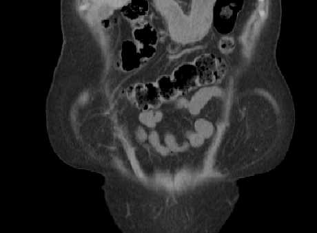
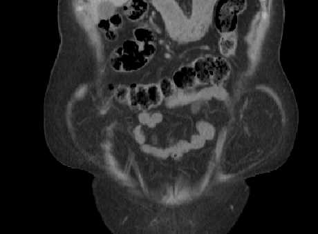
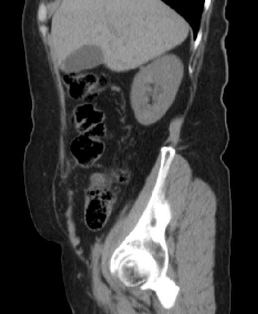
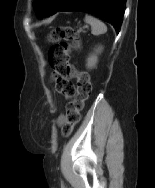
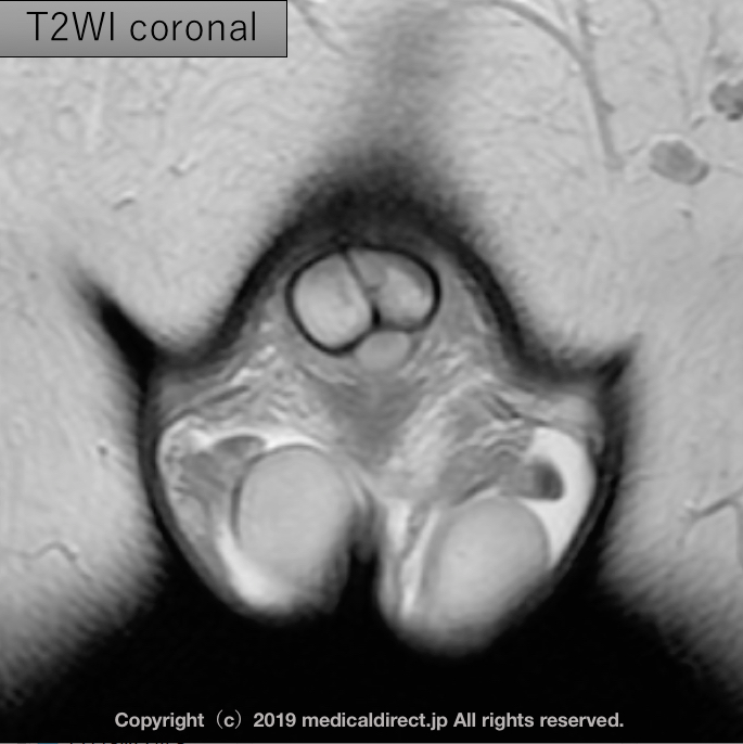
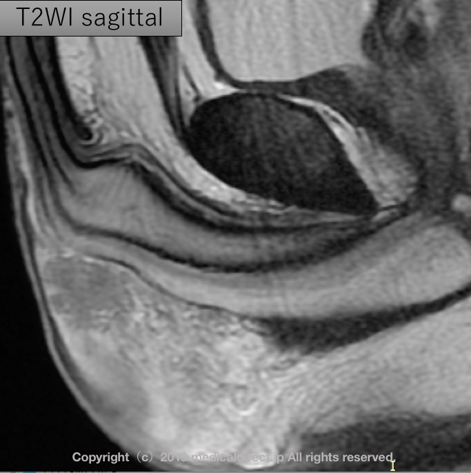
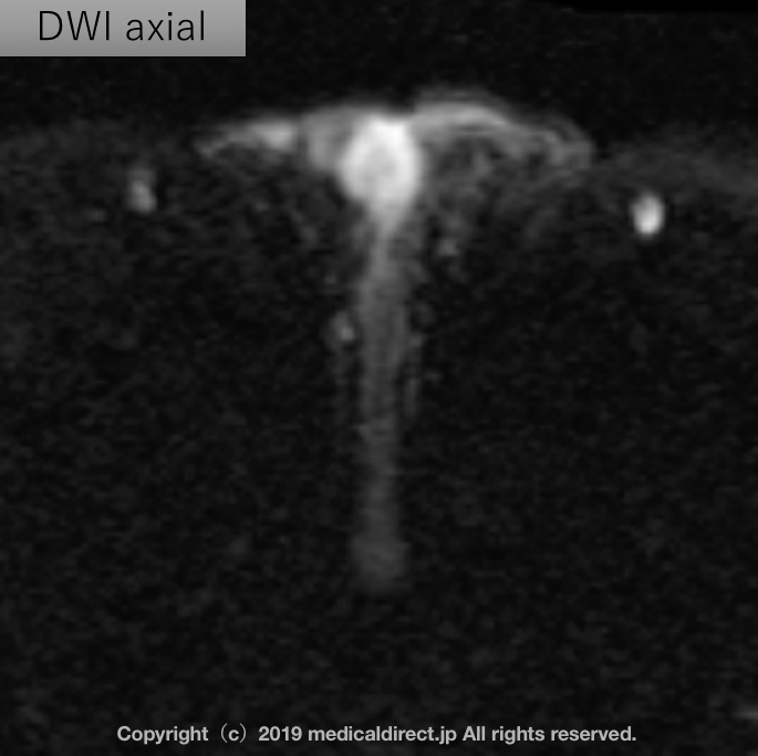
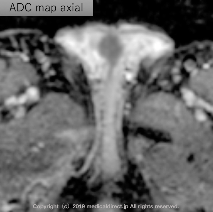
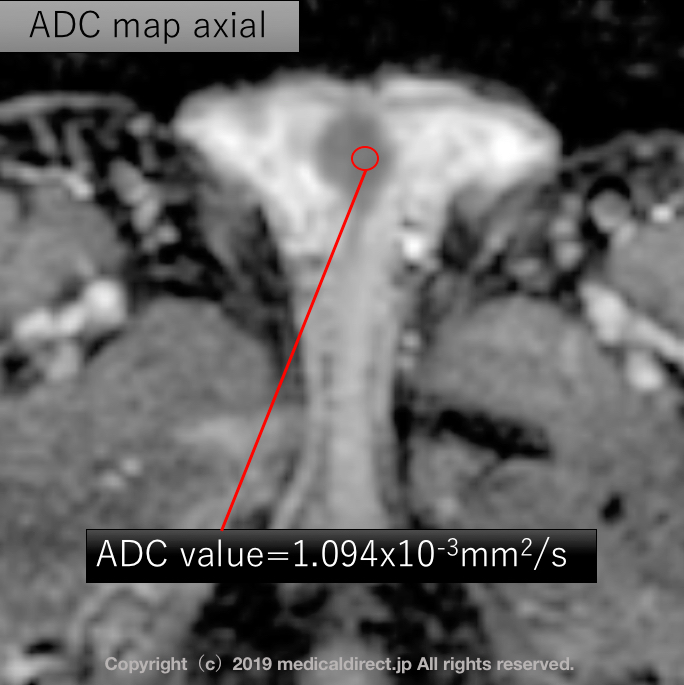
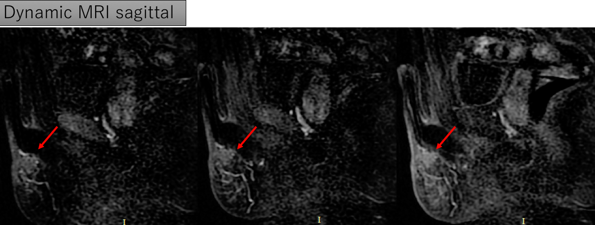
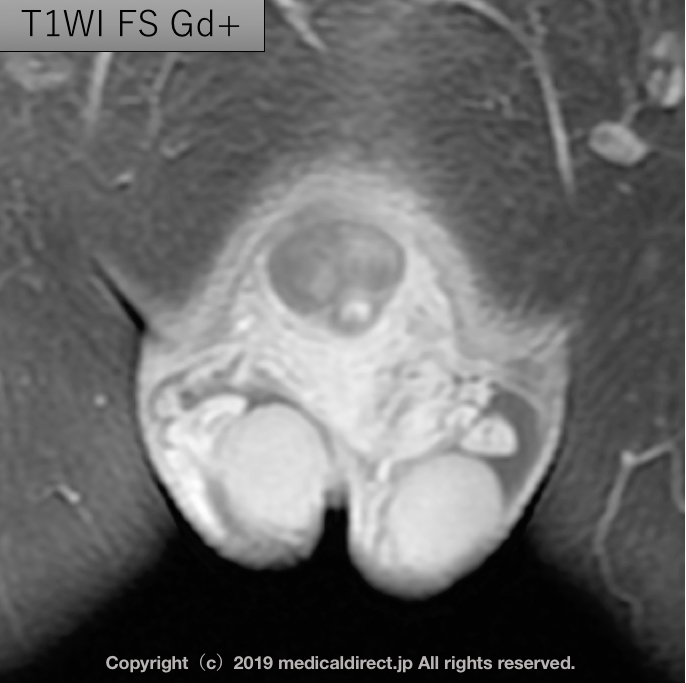
We report the imaging findings of sclerosing lipogranuloma. Sclerosing lipogranuloma is a peculiar granulomatous fatty tissue reaction. The majority of the cases occur in the genital and urinary tracts. To our knowledge, the CT and MR images of this rare entity have not been reported in the English literature. We present a case that was suspected to be sclerosing lipogranuloma of the male genitalia on CT and MR images and was diagnosed by open biopsy.

原発性陰嚢内硬化性脂肪肉芽腫の1例 : 自験例報告と本邦 報告227例の検討

渡邊 大祐, 磯野 誠, 新地 祐介, 他

泌尿器科紀. 2014, 60: 587- 591

https://repository.kulib.kyoto-u.ac.jp/dspace/bitstream/2433/192319/1/60_587.pdf


陰嚢内硬化性脂肪肉芽腫の1例

児玉 浩一, 四柳 智嗣, 布施 春樹, 他

泌尿器科紀要. 1999, 45: 211-214

https://repository.kulib.kyoto-u.ac.jp/dspace/bitstream/2433/114000/1/45_211.pdf


原発性外陰部硬化性脂肪肉芽腫の3例

増田 均, 山田 拓己, 長浜 克志, 他

泌尿器科紀. 1992, 38: 1183-1186

https://repository.kulib.kyoto-u.ac.jp/dspace/bitstream/2433/117667/1/38_1183.pdf


陰嚢内硬化性脂肪肉芽腫の 1例

深堀 能立, 鏑 木 豊, 猿木 和 久, 他

北関東医学.1988. 38: 175~182

https://www.jstage.jst.go.jp/article/kmj1951/38/3/38_3_175/_pdf/-char/en

45-year-old man with vertigo. 45才男性 めまい

45-year-old man with vertigo.
45才男性 めまい

解答:  Ecchordosis physaliphora   (EP)  ,  泡状外脊索腫

iPad version


You tube version

問題を見ていない先生はこちら

Key words: Ecchordosisphysaliphora, EP,  泡状外脊索症,  異所性 脊索の遺残

Key images:

T2WI 3D axial 3

通常のT2WI だとわかりにくい 上記の様に3DT2WI (今回は内耳道撮像目的)でないと難しい

T2WI axialT2WI 3D axi reconstruction sagi, coro ADCT1WI T2WI MPR3T1WI T2WI MPRT1WI T2WI MPR2

References:

1)MehnertF, Beschorner, KukerW,et al.

Retroclivalecchordosisphysaliphora: MR imaging and review of the literature. AJNR. 2004, 25: 1851-1855

2)   ChiharaC, Korogi Y, Kakeda S, et al.

Ecchordosis physaliphora and its variants: proposed new classification based on high-resolution fast MR imaging employing steady-state acquisition. EurRadiol. 2013 Oct;23(10):2854-60.

3) Srinivasan A, Goyal M, KingstoneM.

Case 133:

Ecchordosis physaliphora. Radiology. 2008 May;247(2):585-8. No abstract available.

4) Golden LD, Small JE Benign notochordal lesions of the posterior clivus:      retrospective review of prevalence and imaging characteristics. J Neuroimaging.2014 May-Jun;24(3):245-9.

5) KaulS, Khan OH, Edem I, et al.

Transclivalpseudomeningocele secondary to ecchordosisphysaliphora: case report and literature review. J NeurolSurg Rep. 2013 Dec;74(2):92-5.

6)  KrishtKM, Palmer CA, Osborn AG, et al.

Giant ecchordosis physaliphora in an adolescent girl: case report. J NeurosurgPediatr. 2013 Oct;12(4):328-33. doi: 10.3171/2013.5.PEDS1395. Epub 2013 Aug 2.

7)Yamamoto T, Yano S, Hide T, et al.

A case of ecchordosis physaliphora presenting with an abducens nerve palsy: A rare symptomatic case managed with endoscopic endonasaltranssphenoidalsurgery. SurgNeurol Int. 2013;4:13.

8)AlkanO, Yildirim T, Kizilkiliç O, et al.

A case of ecchordosis physaliphora presenting with an intratumoral hemorrhage. Turk Neurosurg. 2009 Jul;19(3):293-6.

9)CiarpagliniR, Pasquini E, Mazzatenta D, et al.

Intraduralclivalchordoma and ecchordosis physaliphora: a challenging differential diagnosis: case report. Neurosurgery. 2009 Feb;64(2):E387-8; discussion E388.

10) Toda H, Kondo A, Iwasaki K.

Neuroradiologicalcharacteristics of ecchordosis physaliphora. Case report and review of the literature. J Neurosurg. 1998 Nov;89(5):830-4. Review.

55-year-old woman with vertigo. Answer 解答編

55-year-old woman with vertigo. 解答編

問題を見ていない先生はまず問題から

iPad version はこちら

You tube version はこちら

Key words: 巨大くも膜顆粒, くも膜顆粒、giant arachnoid granulation, arachnoid granulation, AG, AGs

Key comment: AGs  くも膜顆粒とは?

  • 通常1cm 未満:2-8mm、
  • 古いAJNRの論文だとCT上24%, MRI上13%で(造影MRIの静脈洞の濃染欠損域として)認められる 。平均的大きさは4-5mm
  • ほとんどが画像的に横静脈洞にみられる
  • 次に多いのが静脈洞交会(TorcularHerophili)
  • 剖検例29例も検討し類似結果を得ている 出現頻度 66%
  • 剖検では1-8mmで平均2mm
  • 性差はない
  • くも膜顆粒が存在する群は有意に年齢が高め
  • 無症状がほとんどで偶然見つかる、稀に頭痛

巨大なくも膜顆粒 って? giant AGs

  • 巨大くも膜顆粒:1cm を超えるもの
  • 19個の1cmを超えるくも膜顆粒を検討した45-75才 17人(2人が2つのAGs有す)
  • 2/17症例 頭痛あり
  • 横静脈洞:12、上矢状洞:6、静脈洞交会:1
  • CSF と同等の信号域と言われていたが・・・80%はCSFと同等ではない 何故?
  • AGsは単純にCSFの袋ではない→ ビデオを見てみてくださいね。

References:

1) Leach JL, Jones BV,  Tomsick TA, et al. Normal appearance of arachnoid granulations on contrast-enhanced CT and MR of the brain: differentiation from dural sinus disease.

AJNR Am J Neuroradiol.1996 Sep;17(8):1523-32.

2)  Trimble CR, Harnsberger HR, Castillo M, Brant-Zawadzki M, Osborn AG.

“Giant” arachnoid granulations just like CSF?: NOT!!

AJNR Am J Neuroradiol. 2010 Oct;31(9):1724-8. doi: 10.3174/ajnr.A2157.


55-year-old woman with vertigo. めまいを主訴に来院された55才女性

56才女性 めまいを主訴に来院

先生がこの画像を見て手はとまりますか? すんなり読影しますか?

iPad version.

You tube version

解答は今年度内に予定

Abdominal wall hernia Answer 解答編

Abdominal wall hernia: Spiegelian hernia  半月状線ヘルニア

問題をみていないので、問題を見てからこの回答を見る

iPad version


You tube version

key words: Abdominal wall hernia, 腹壁ヘルニア、Spiegelian hernia, 半月状線ヘルニア、hernia, ヘルニア、surgery, 手術

Spiegelian hernia まとめ

  • 半月状線ヘルニア:腹横筋線維が腱膜に移行する腹直筋外縁の半月状線,すなわちSpigel腱膜に発生する稀なヘルニア
  • 半月状線の脆弱性、上前腸骨棘を結ぶ線から6cm上方までに発生しやすい=SH belt
  • 平均年齢60才台、女性優位
  • 左優位、両側性は稀
  • 修復術を行った腹壁ヘルニア中の頻度は1%未満
  • 原因として外科手術や極度の肥満など
  • 外腹斜筋腱膜をこえるか否かで2つに分類

–Interstitial SH: closed loop Small bowel obstruction なり易い

–Subcutaneous SH

  • CTで100%の感度とPPV.

Key images

半月状線とはどこ?1半月状線とはどこ?2

CT0050CT0056

CT0009CT0010CT0011CT0012CT0015CT0048文献:

1.Martine M, Paquette B, Badet N, et al. Spiegelian hernia: CT findings and clinical relevance. Abdom imaging. 2013; 38: 260-264

2. Moles Morenilla L, DocoboDurántez F, Mena Robles J,etal. Spiegelianhernia in Spain. An analysis of 162 cases. Rev EspEnferm Dig. 2005 May;97(5):338-47.

3. Light D, Chattopadhyay D, Bawa S. Radiological and clinical examination in the diagnosis of Spiegelian hernia. Ann R CollSurgEngl 2013; 95:98-10

4. Dabbas N, Adams K, Pearson K, et al.  Frequency of abdominal wall hernias: is classical teaching out of date?   JRSM Short Rep.2011 Jan 19;2(1):5. doi: 10.1258/shorts.2010.010071.

5.  塩田喜代美、植木孝宣、青井重善、他。CTにて術前診断した半月状線ヘルニアの1例  日臨外会誌 . 2002: 63: 1308-1311

English summary

Summary of Spiegelian hernia

  • Spigelian hernias (SH) are rarely reported abdominal wallhernias, which arise from the Spigelianfascia. It lies along the semilunar line lateral to the rectus abdominis muscle.
  • SH is due to a weakness of the spiegelian fascia.
  • Most of the SH cases occur within the zone, so-called SH belt, which is a transverse band between the line joining both anterior superior iliac spines and parallel line lying 6-cm cranial to it.
  • Mean age is 60-70 year-old. Female domination.
  • Left side is dominant. Bilateral SH is very rare (2%).
  • The incidence of SH among abdominal wall hernia repair surgery is less than 1%.
  • Major acquired risk factors of SH are a history of abdominal surgery and obesity.
  • Two types of SH are interstitial SH and subcutaneous SH. Closed loop small bowel obstruction is statistically associated with interstitial SH.
  • CT has a 100% sensitivity and 100% PPV todiagnose SH.