44-year-old man with vertigo. Answer.
Tornwaldt cyts
iPad version
WMV version
key words: Tornwaldt cyst, トーンワルト嚢胞, mucous retention cyst
44-year-old man with vertigo. Answer.
Tornwaldt cyts
iPad version
WMV version
key words: Tornwaldt cyst, トーンワルト嚢胞, mucous retention cyst
45-year-old man with vertigo.
45才男性 めまい
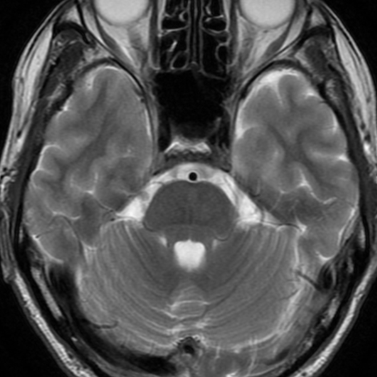
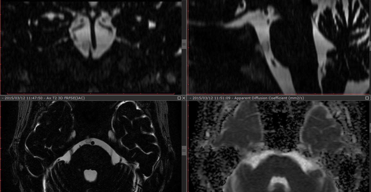
解答: Ecchordosis physaliphora (EP) , 泡状外脊索腫
iPad version
You tube version
Key words: Ecchordosisphysaliphora, EP, 泡状外脊索症, 異所性 脊索の遺残
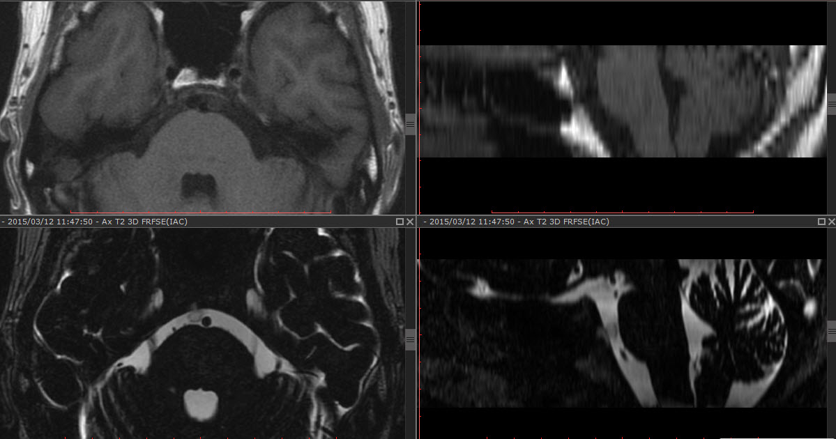
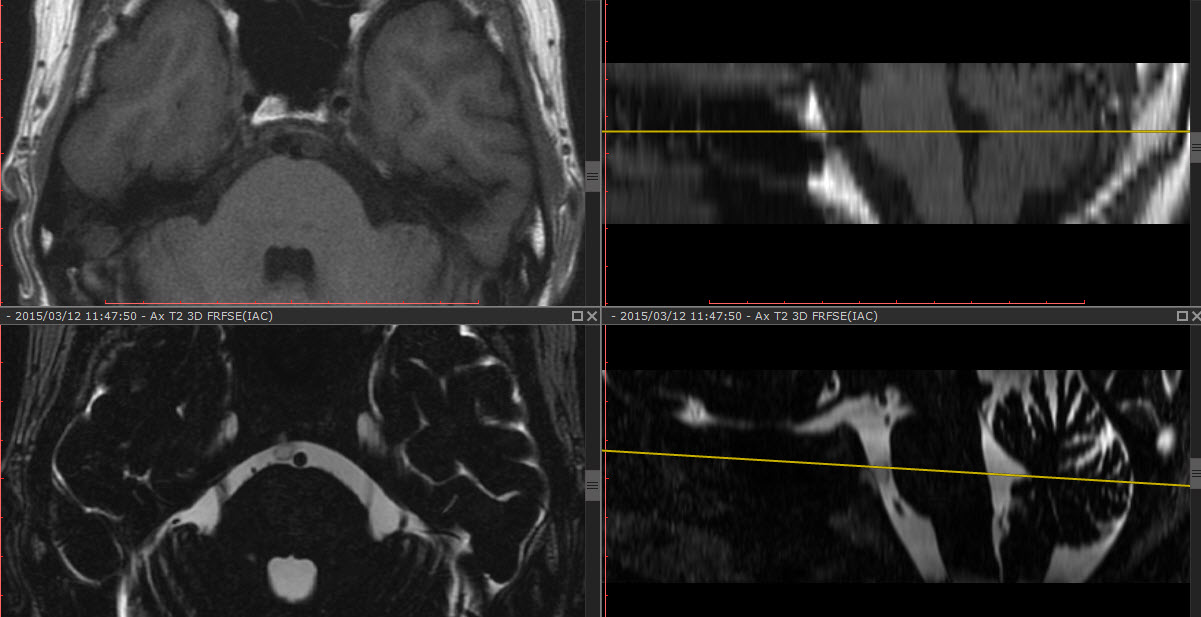
Key images:
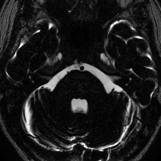
通常のT2WI だとわかりにくい 上記の様に3DT2WI (今回は内耳道撮像目的)でないと難しい

References:
1)MehnertF, Beschorner, KukerW,et al.
Retroclivalecchordosisphysaliphora: MR imaging and review of the literature. AJNR. 2004, 25: 1851-1855
2) ChiharaC, Korogi Y, Kakeda S, et al.
Ecchordosis physaliphora and its variants: proposed new classification based on high-resolution fast MR imaging employing steady-state acquisition. EurRadiol. 2013 Oct;23(10):2854-60.
3) Srinivasan A, Goyal M, KingstoneM.
Case 133:
Ecchordosis physaliphora. Radiology. 2008 May;247(2):585-8. No abstract available.
4) Golden LD, Small JE Benign notochordal lesions of the posterior clivus: retrospective review of prevalence and imaging characteristics. J Neuroimaging.2014 May-Jun;24(3):245-9.
5) KaulS, Khan OH, Edem I, et al.
Transclivalpseudomeningocele secondary to ecchordosisphysaliphora: case report and literature review. J NeurolSurg Rep. 2013 Dec;74(2):92-5.
6) KrishtKM, Palmer CA, Osborn AG, et al.
Giant ecchordosis physaliphora in an adolescent girl: case report. J NeurosurgPediatr. 2013 Oct;12(4):328-33. doi: 10.3171/2013.5.PEDS1395. Epub 2013 Aug 2.
7)Yamamoto T, Yano S, Hide T, et al.
A case of ecchordosis physaliphora presenting with an abducens nerve palsy: A rare symptomatic case managed with endoscopic endonasaltranssphenoidalsurgery. SurgNeurol Int. 2013;4:13.
8)AlkanO, Yildirim T, Kizilkiliç O, et al.
A case of ecchordosis physaliphora presenting with an intratumoral hemorrhage. Turk Neurosurg. 2009 Jul;19(3):293-6.
9)CiarpagliniR, Pasquini E, Mazzatenta D, et al.
Intraduralclivalchordoma and ecchordosis physaliphora: a challenging differential diagnosis: case report. Neurosurgery. 2009 Feb;64(2):E387-8; discussion E388.
10) Toda H, Kondo A, Iwasaki K.
Neuroradiologicalcharacteristics of ecchordosis physaliphora. Case report and review of the literature. J Neurosurg. 1998 Nov;89(5):830-4. Review.
55-year-old woman with vertigo. 解答編
iPad version はこちら
You tube version はこちら
Key words: 巨大くも膜顆粒, くも膜顆粒、giant arachnoid granulation, arachnoid granulation, AG, AGs
Key comment: AGs くも膜顆粒とは?
巨大なくも膜顆粒 って? giant AGs
References:
1) Leach JL, Jones BV, Tomsick TA, et al. Normal appearance of arachnoid granulations on contrast-enhanced CT and MR of the brain: differentiation from dural sinus disease.
AJNR Am J Neuroradiol.1996 Sep;17(8):1523-32.
2) Trimble CR, Harnsberger HR, Castillo M, Brant-Zawadzki M, Osborn AG.
“Giant” arachnoid granulations just like CSF?: NOT!!
AJNR Am J Neuroradiol. 2010 Oct;31(9):1724-8. doi: 10.3174/ajnr.A2157.