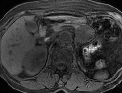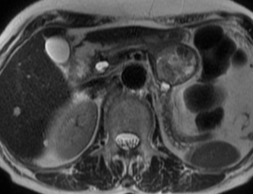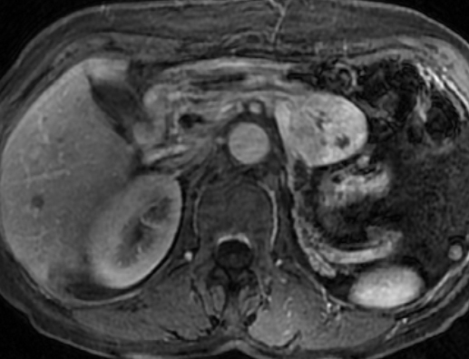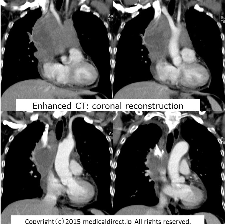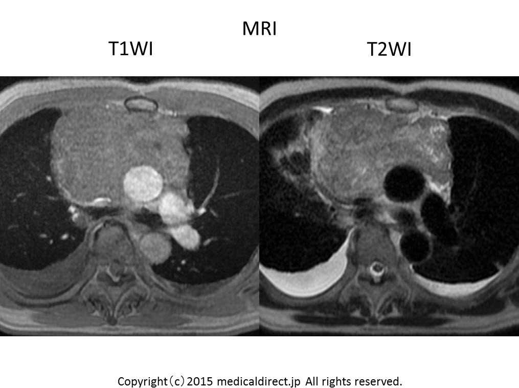
74才男性
超音波スクリーニングにて
膵尾部腫瘍を指摘
74-year-old man with tumor in pancreatic tail on screening US.
解答編
iPad version
WMV version
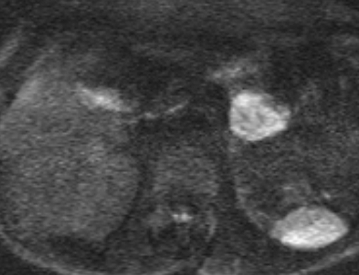
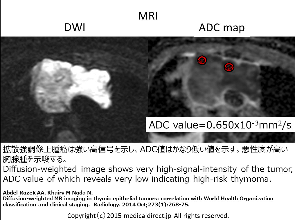
Key words: Pancreatic metastases, 膵転移、Renal cell carcinoma, RCC, 腎細胞癌、腎癌、腎癌膵転移、RCC, diffusion, ADC map
Key images:


Referrences:
Clinical features, Prognosis
1. Song SW, Cheng JF, Liu N, Zhao TH.
Diagnosis and treatment of pancreatic metastases in 22 patients: a retrospective study.WorldJ SurgOncol. 2014 Sep 25;12:299.
2. SellnerF, Tykalsky N, De Santis M, et al.
Solitary and multiple isolated metastases of clear cell renal carcinoma to thepancreas: an indication for pancreatic surgery.
Ann SurgOncol. 2006 Jan;13(1):75-85. Epub 2006 Jan 1.
3. ZerbiA, Ortolano E, Balzano G, et al.
Pancreatic metastasis from renal cell carcinoma: which patients benefit from surgical resection?Ann SurgOncol. 2008 Apr;15(4):1161-8.
4. Law CH, Wei AC, Hanna SS, et al.
Pancreatic resection for metastatic renal cell carcinoma: presentation, treatment, and outcome. Ann Surg Oncol.2003 Oct;10(8):922-6.
5. Sohn TA, Yeo CJ, Cameron JL, et al.
Renal cell carcinoma metastatic to the pancreas: results of surgical management.J GastrointestSurg.2001 Jul-Aug;5(4):346-51.
6. Faure JP, Tuech JJ, Richer JP, et al.
Pancreatic metastasis of renal cell carcinoma: presentation, treatment and survival. J Urol. 2001 Jan;165(1):20-2.
7. AkatsuT, Shimazu M, Aiura K, et al.
Clinicopathological features and surgical outcome of isolated metastasis of renal cell carcinoma. Hepatogastroenterology. 2007 Sep;54(78):1836-40.
Imaging features
1. Ng CS, Loyer EM, Iyer RB, David CL, DuBrow RA, Charnsangavej C.
Metastases to the pancreas from renal cell carcinoma: findings on three-phase contrast-enhanced helical CT. AJRAm J Roentgenol. 1999 Jun;172(6):1555-9.
2. Klein KA, Stephens DH, Welch TJ.
CT characteristics of metastatic disease of the pancreas. Radiographics. 1998 Mar-Apr;18(2):369-78.
3. VincenziM, Pasquotti G, Polverosi R, et al.
Imaging of pancreatic metastases from renal cell carcinoma. Cancer Imaging. 2014 Apr 22;14:5.
4. PalmowskiM, Hacke N, Satzl S, et al.
Metastasis to the pancreas: characterization by morphology and contrast enhancement features on CT and MRI. Pancreatology. 2008;8(2):199-203.
5. AscentiG, Visalli C, Genitori A, et al.
Multiple hypervascular pancreatic metastases from renal cell carcinoma: dynamic MR and spiral CT in three cases. Clin Imaging. 2004 Sep-Oct;28(5):349-52.
6. Tsitouridis I, Diamantopoulou A, Michaelides M, et al.
Pancreatic metastases: CT and MRI findings. DiagnIntervRadiol. 2010 Mar;16(1):45-51.
RCC and ADC value
1. Choi YA, Kim CK, Park SY, et al.
Subtype differentiation of renal cell carcinoma using diffusion-weighted and blood oxygenation level-dependent MRI.AJR Am J Roentgenol. 2014 Jul;203(1):W78-84.
2. SasamoriH, Saiki M, Suyama J, et al.
Utility of apparent diffusion coefficients in the evaluation of solid renal tumors at 3T. MagnReson Med Sci. 2014;13(2):89-95.
3. Sevcenco S, Heinz-Peer G, Ponhold L, et al.
Utility and limitations of 3-Tesla diffusion-weighted magnetic resonance imaging for differentiation of renal tumors. EurJ Radiol. 2014 Jun;83(6):909-13.
4. Yu X, Lin M, Ouyang H, et al.
Application of ADC measurement in characterization of renal cell carcinomas with different pathological types and grades by 3.0T diffusion-weighted MRI.
EurJ Radiol. 2012 Nov;81(11):3061-6.
5. PaudyalB, Paudyal P, Tsushima Y, et al.
The role of the ADC value in the characterisation of renal carcinoma by diffusion-weighted MRI. Br J Radiol. 2010 Apr;83(988):336-43.
RCC and PET-CT
1. Win AZ, Aparici CM.
Clinical effectiveness of (18)f-fluorodeoxyglucose positron emission tomography/computed tomography in management of renal cell carcinoma: a single institution experience. World J Nucl Med. 2015 Jan-Apr;14(1):36-40.
2. Sharma P, Karunanithi S, Chakraborty PS, et al.
18F-Fluoride PET/CT for detection of bone metastasis in patients with renal cell carcinoma: a pilot study. NuclMed Commun. 2014 Dec;35(12):1247-53.
3. Lee H, Hwang KH, Kim SG, et al.
Can Initial (18)F-FDG PET-CT Imaging Give Information on Metastasis in Patients with Primary Renal Cell Carcinoma? NuclMed Mol Imaging. 2014 Jun;48(2):144-52.
4. Fuccio C, Ceci F, Castellucci P, et al.
Restaging clear cell renal carcinoma with 18F-FDG PET/CT. ClinNucl Med. 2014 Jun;39(6):e320-4.
5. Chen JL, Appelbaum DE, Kocherginsky M, et al.
FDG-PET as a predictive biomarker for therapy with everolimus in metastatic renal cell cancer. Cancer Med. 2013 Aug;2(4):545-52.
6. Nakhoda Z, Torigian DA, Saboury B, et al.
Assessment of the diagnostic performance of (18)F-FDG-PET/CT for detection and characterization of solid renal malignancies. Hell J Nucl Med. 2013 Jan-Apr;16(1):19-24.
7. Wang HY, Ding HJ, Chen JH, et al.
Meta-analysis of the diagnostic performance of [18F]FDG-PETand PET/CT in renal cell carcinoma. Cancer Imaging. 2012 Oct 26;12:464-74.
8. Namura K, Minamimoto R, Yao M, et al.
Impact of maximum standardized uptake value (SUVmax) evaluated by 18-Fluoro-2-deoxy-D-glucose positron emission tomography/computed tomography (18F-FDG-PET/CT) on survival for patients with advanced renal cell carcinoma: a preliminary report. BMC Cancer. 2010 Dec 3;10:667.
9. Mueller-Lisse UG, Mueller-Lisse UL.
Imaging of advanced renal cell carcinoma. World J Urol. 2010 Jun;28(3):253-61.
10. Rodríguez Martínez de Llano S, Jiménez-Vicioso A, et al.
Clinical impact of (18)F-FDG PET in management of patients with renal cell carcinoma.
Rev Esp Med Nucl. 2010 Jan-Feb;29(1):12-9.
11. Staging of renal cell carcinoma.
Mueller-Lisse UG, Mueller-Lisse UL, Meindl T, et al. EurRadiol. 2007 Sep;17(9):2268-77.
12. Ak I, Can C.
F-18 FDG PET in detecting renal cell carcinoma. ActaRadiol. 2005 Dec;46(8):895-9.