iphone ver
タグ別アーカイブ: liver
保護中: 文献カンファランスシリーズ 第5回(メルマガ購読者のみ再生できます)
EOB プリモビスト症例集 第7回 回答
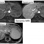
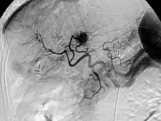
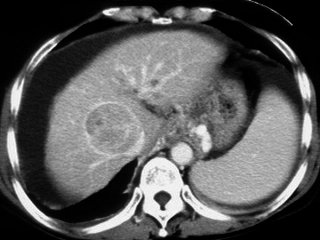
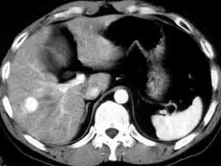
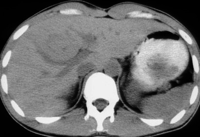
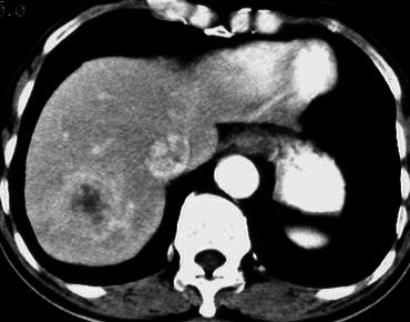
key words: high flow hemangioma, atypical hemangioma, EOB primovist MRI
イチロウ サイト内の関連ページ
肝画像診断 症例6 の動画参照
http://medicaldirect.jp/shourei6/kaitou62.html
<参考文献>
High-flow hemangioma について
Kim KW, Kim AY, Kim TK, et al.
Hepatic hemangiomas with arterioportal shunt: sonographic appearances with CT and MRI correlation.
AJR Am J Roentgenol. 2006 Oct;187(4):W406-14.
http://www.ncbi.nlm.nih.gov/pubmed/16985113
Jang HJ, Kim TK, Lim HK, et al.
Hepatic hemangioma: atypical appearances on CT, MR imaging, and sonography.
AJR, 2003 Jan;180(1):135-41.
http://www.ncbi.nlm.nih.gov/pubmed/12490492
Vilgrain V, Boulos L, Vullierme MP,et al.
Imaging of atypical hemangiomas of the liver with pathologic correlation.
Radiographics. 2000 Mar-Apr;20(2):379-97.
http://www.ncbi.nlm.nih.gov/pubmed/10715338
Kato H, Kanematsu M, Matsuo M, et al.
Atypically enhancing hepatic cavernous hemangiomas: high-spatial-resolution gadolinium-enhanced triphasic dynamic gradient-recalled-echo imaging findings.
Eur Radiol. 2001;11(12):2510-5. Epub 2001 Sep 7.
Yan FH, Zeng MS, Zhou KR
Role and pitfalls of hepatic helical multi-phase CT scanning in differential diagnosis of small hemangioma and small hepatocellular carcinoma.
World J Gastroenterol 1998 Aug;4(4):343-347.
Yu JS, Kim KW, Park MS, Yoon SW.
Hepatic cavernous hemangioma in cirrhotic liver: imaging findings.
Korean J Radiol. 2000 Oct-Dec;1(4):185-90.
EOB プリモビスト症例集 第5回 回答
参考文献
*dysplastic nodules 文献
Earls JP, et al.
Dysplastic nodules and hepatocellular carcinoma: thin-section MR imaging of explanted cirrhotic livers with pathologic correlation.
Radiology 1996 Oct;201(1):207-14.
http://www.ncbi.nlm.nih.gov/pubmed/8816545
Early hepatocelluar carcinoma: MR imaging
Muramatsu Y, et al.
Radiology. 1991 Oct;181(1):209-13.
http://www.ncbi.nlm.nih.gov/pubmed/1653443
Matsui O, et al.
Adenmatous hperplastic nodules in the cirrhotic liver: differentiation from hepatocellular carcinoma with MR imaging.
Radiology. 1989 Oct;173(1):123-6.
http://www.ncbi.nlm.nih.gov/pubmed/2550995
*最新の論文: T1WI 高信号を呈する結節のEOB primovist診断
Chou CT, et al. Characterization of hyperintense nodules on precontrast T1-weighted MRI: utility of gadoxetic acid-enhanced hepatocyte-phase imag
J Magn Reson Imaging. 2011 Mar;33(3):625-32.
http://www.ncbi.nlm.nih.gov/pubmed/21563246
*結節の分化度と肝細胞相での信号強度の相関性を検討した論文
Kogita S, Imai Y, Okada M, et al. Gd-EOB-DTPA-enhanced magnetic resonance images of hepatocellular carcinoma: correlation with histological grading and portal blood flow.
Eur Radiol. 2010 Oct;20(10):2405-13. Epub 2010 May 19.
肝臓CT、MRI 症例集(随時更新)
症例1 50歳男性、糖原病(画像をクリック)
症例2 71歳男性、C型肝硬変(画像をクリック)
症例3 55歳女性(画像をクリック)
症例4 58歳男性、B型肝硬変(画像をクリック)
症例5 60歳女性、肝硬変(画像をクリック)
症例6 40歳男性、検診にて指摘(画像をクリック)
症例7 50歳女性、検診にて指摘(画像をクリック)
症例8 50歳女性、検診にて肝腫瘤指摘(画像をクリック)
症例9 21歳男性、検診にて腫瘤を指摘(画像をクリック)
症例10 60歳男性(画像をクリック)
症例11 55歳 男性
症例12 60歳 男性
症例13
症例14(画像をクリック)
エキスパートはこう考える第2回 回答
iphone用動画
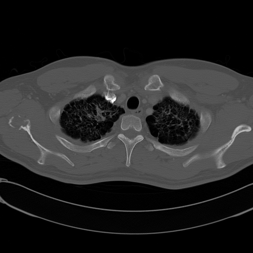
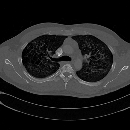
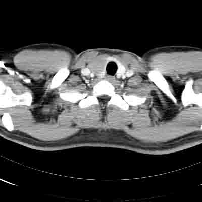
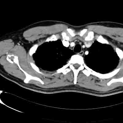
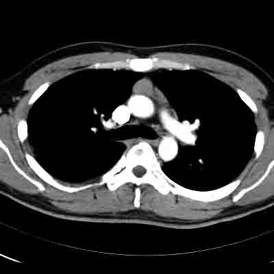
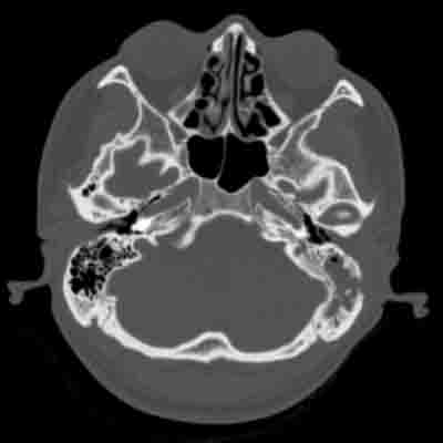
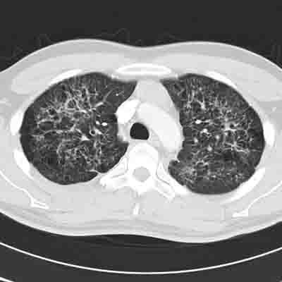
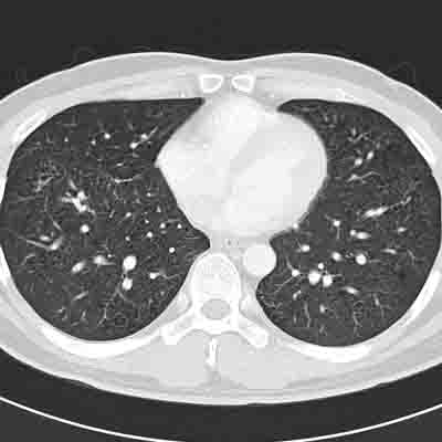
<key word>喫煙、尿崩症、両肺対称性網状陰影、上肺優位の縮み、多発空洞性陰影、多発結節影、骨病変、Langerhans cell histiocytosis, LCH, eosinophilic granuloma, 好酸球性肉芽腫
<key画像>
 下垂体 MRI 下垂体 MRI |
骨病変 1 |
|
骨病変 2 |
縦隔条件 1 |
|
縦隔条件 2 |
縦隔条件 3 |
|
側頭骨 1 |
肺野条件 1 |
 肺野条件 2 肺野条件 2 |
肺野条件 3 |
<文献>
LCH肺
Abbott GF, Rosado-de-Christenson ML, et al.
From the archives of the AFIP: pulmonary Langerhans cell
histiocytosis.
Radiographics. 2004 May-Jun;24(3):821-41.
http://radiographics.rsna.org/content/24/3/821.full.pdf+html
Leatherwood DL, Heitkamp DE, Emerson RE.
Best cases from the AFIP: Pulmonary Langerhans cell
histiocytosis.
Radiographics. 2007 Jan-Feb;27(1):265-8.
http://radiographics.rsna.org/content/27/1/265.full.pdf+html
Sundar KM, Gosselin MV, Chung HL, Cahill BC.
Pulmonary Langerhans cell histiocytosis: emerging concepts
in pathobiology, radiology, and clinical evolution of
disease.
Chest. 2003 May;123(5):1673-83.
http://chestjournal.chestpubs.org/content/123/5/1673.full.pdf+html
Kulwiec EL, Lynch DA, Aguayo SM, Schwarz MI, King TE Jr.
Imaging of pulmonary histiocytosis X.
Radiographics. 1992 May;12(3):515-26.
http://radiographics.rsna.org/content/12/3/515.long
LCH縦隔
Donnelly LF, Frush DP.
Langerhans’ cell histiocytosis showing low-attenuation
mediastinal mass and cystic lung disease.
AJR Am J Roentgenol. 2000 Mar;174(3):877-8.
http://www.ajronline.org/cgi/content/full/174/3/877-a
LCH 全般
Meyer JS, Harty MP, Mahboubi S, Heyman S, Zimmerman RA,
Womer RB, Dormans JP, D’Angio GJ.
Langerhans cell histiocytosis: presentation and evolution
of radiologic findings with clinical correlation.
Radiographics. 1995 Sep;15(5):1135-46.
http://radiographics.rsna.org/content/15/5/1135.long
LCH 頭部
D’Ambrosio N, Soohoo S, Warshall C, Johnson A, Karimi S.
Craniofacial and intracranial manifestations of langerhans
cell histiocytosis: report of findings in 100 patients.
AJR Am J Roentgenol. 2008 Aug;191(2):589-97.
http://www.ajronline.org/cgi/reprint/191/2/589
Grois N, Prayer D, Prosch H, Lassmann H; CNS LCH
Co-operative Group.
Neuropathology of CNS disease in Langerhans cell
histiocytosis.
Brain. 2005 Apr;128(Pt 4):829-38. Epub 2005 Feb 10.
http://brain.oxfordjournals.org/cgi/reprint/128/4/829
Prayer D, Grois N, Prosch H, Gadner H, Barkovich AJ.
MR imaging presentation of intracranial disease associated
with Langerhans cell histiocytosis.
AJNR Am J Neuroradiol. 2004 May;25(5):880-91.
http://www.ajnr.org/cgi/reprint/25/5/880
LCH骨
Stull MA, Kransdorf MJ, Devaney KO.
Langerhans cell histiocytosis of bone.
Radiographics. 1992 Jul;12(4):801-23.
http://radiographics.rsna.org/content/12/4/801.long
Sartoris DJ, Parker BR.
Histiocytosis X: rate and pattern of resolution of osseous
lesions.
Radiology. 1984 Sep;152(3):679-84.
http://radiology.rsna.org/content/152/3/679.long
LCH甲状腺
Uchiyama M, Watanabe R, Ito I, Ikeda T.
Thyroid involvement in pulmonary langerhans cell
histiocytosis.
Intern Med. 2009;48(23):2047-8. Epub 2009 Dec 1.
http://www.jstage.jst.go.jp/article/internalmedicine/48/23/2047/_pdf
LCH肝
Radin DR.
Langerhans cell histiocytosis of the liver: imaging
findings.
AJR Am J Roentgenol. 1992 Jul;159(1):63-4.

