


文献:
急性膵炎診療ガイドライン 2010年
http://www.suizou.org/etc.htm よりダウンロードを。
一般情報:難病情報セン ター ホームページへのリンク
http://www.nanbyou.or.jp/entry/271
Balthazal EJ.
Acute Pancreatitis: assessment of severity with clinical and CT evaluation.
Radiology 2002; 223:603-613
のp 608 より引用抜粋改変
CT Enhancement Values and Pitfalls
With helical or multidetector scanning with rapid acquisition of sequential images and collimation of less than 5 mm, images can be obtained in the early portal venous phase (60.70 seconds after intravenous administration of 150 mL of iodinated contrast material at a rate of 3 mL/sec). We still use a monophasic protocol, starting at the top of the diaphragm and covering the entire abdomen and pelvis. Since unenhanced images are not obtained, the detection of parenchymal injury is based solely on the degree and homogeneity of pancreatic enhancement. Basic pancreatic CT numbers of 40 .50 HU seen on unenhanced CT images are expected to increase to 100–150 HU throughout the entire normal gland during contrast material administration, depending on the size of the bolus, the speed of the injection, and the time of image acquisition (Fig 3). Lack of contrast enhancement or minimal contrast enhancement of less than 30 HU of a portion of the pancreas or of the entire pancreas indicates decreased blood perfusion (ischemia) and correlates with the development of necrosis (Figs 1, 4, 6). In this regard, however, several factors and potential pitfalls should be kept in mind.
この論文の中で壊死と浮腫をしっかり分けている。かなり具体的:p608-609 CT enhancement value
MDCT 5mm slice 厚、60-70s 3ml/s 150ml 総量
単純CT上膵実質は40-50HUを示す。上記相では、100-150HUまで上昇する
門脈相(60-70s 後)30HU未満では虚血、壊死を示唆する
しかし、いくつかの要因やピットフォールもある。例えば、元もと脂肪変性強い膵の場合、浮腫の強い場合、正常でも そう言う場合がある
もう一つ時間的要素
膵壊死は、臨床症状発症から24-48時間後に生じる。もし、CTが12時間以内に撮影される とよくわからない。はっきりと壊死として濃染しない状態と成るには2-3日かかる。従って、72時間後 の方が壊死の正診率は高まる。48-72時間後がいい
上記もととなったと考えられる膵壊死と病理の対比論文
Larvin M, Chalmers AG, McMahon MJ.
Dynamic contrast enhanced computed tomography: a precise technique for identifying and localising pancreatic necrosis.
BMJ 1990 Jun 2;300(6737):1425-8.
最近の膵壊死のない急性膵炎の予後についての論文
Lenhart DK, Balthazar EJ.
MDCT of acute mild (nonnecrotizing) pancreatitis: abdominal complications and fate of fluid collections.
AJR Am J Roentgenol. 2008 Mar;190(3):643-9.
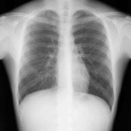
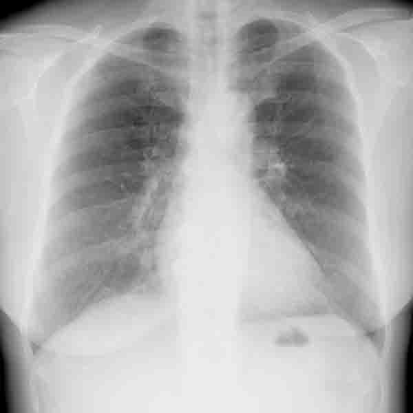
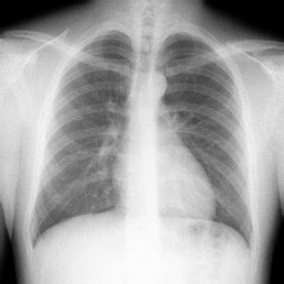
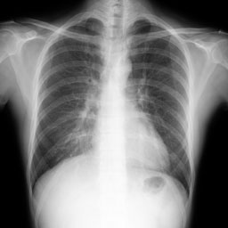
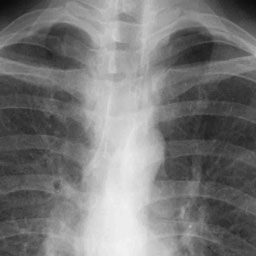
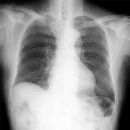 1 年前正常時胸部単純写真
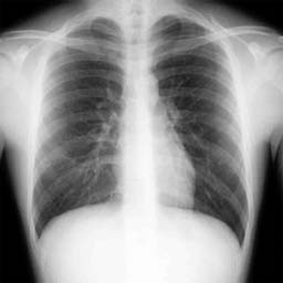
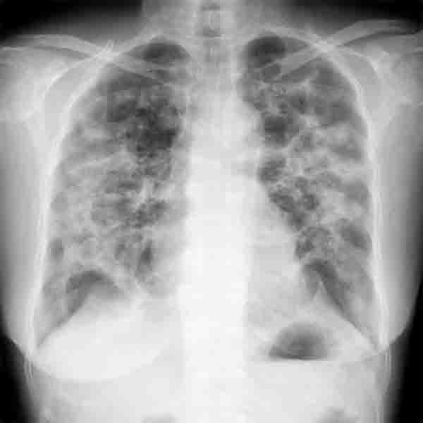
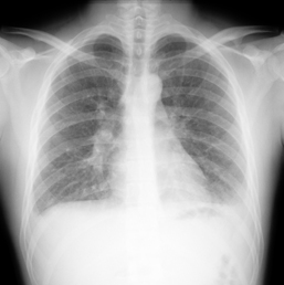
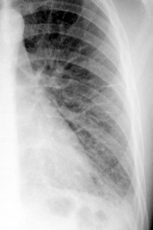
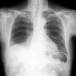 10 ヶ月前肺炎発症時
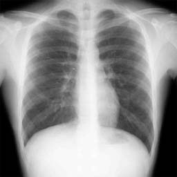
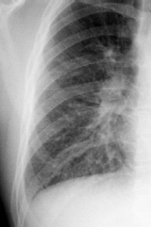
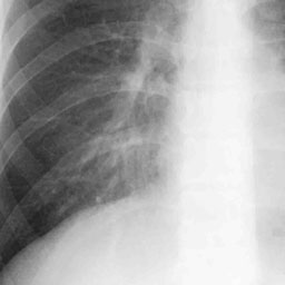
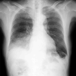 今回の胸部単純写真正面像
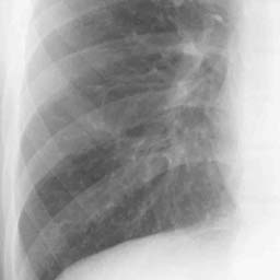
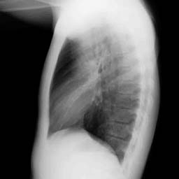
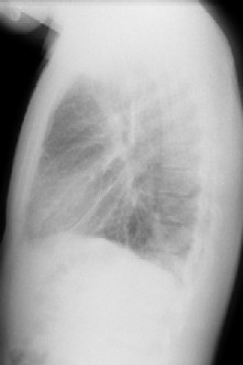
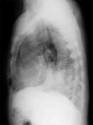 今回の胸部単純写真側面像
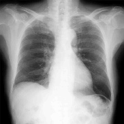 肺炎改善後
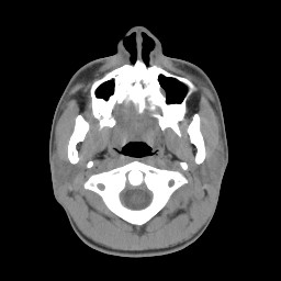
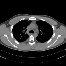
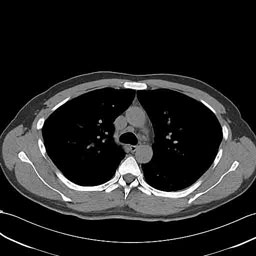
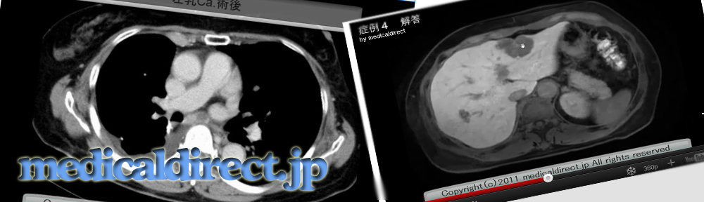
肺炎改善後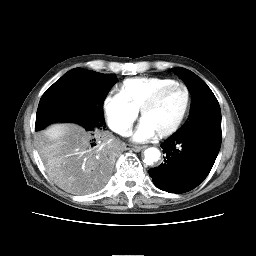 造影 CT 縦隔条件
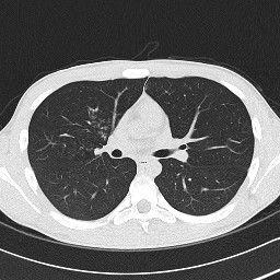
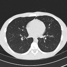
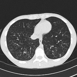
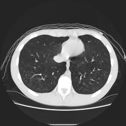
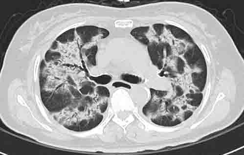
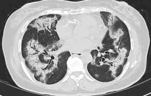
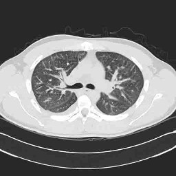
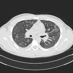
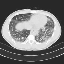
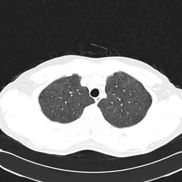
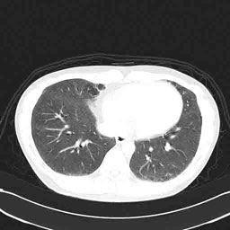
 造影 CT 肺野条件