さて、異常は分かったと思いますが、
先生はその原因はわかります?
3人の放射線科が答えられませんでした
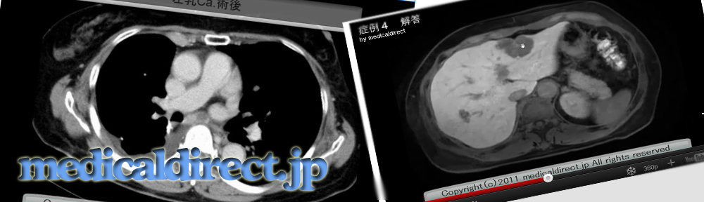


骨シンチグラフィ:向かって左が術前、右が術後

CT:左乳腺B/D領域に乳癌あり、術前に骨シンチグラフィを施行
すると、、、
両側乳房にRI集積が、、、
骨シンチグラフィでRIが乳房に集積するバリエーション
1.大きな乳房:この症例
2.授乳中
3.乳癌
key: 骨シンチグラフィなのに乳房にRI集積、骨シンチ、骨シンチグラフィ、乳房集積、乳房RI集積
key words: 漿液性嚢胞腺腫、漿液性嚢胞腺癌、膵囊胞性病変
Serous cystadenoma, SCA, SCAs, cystic lesions of the pancreas
・SCA の病理と対比したMDCTの診断
Shah AA, Sainani NI, Kambadakone AR, et al.
Predictive value of multi-detector computed tomography for accurate diagnosis of serous cystadenoma: radiologic-pathologic correlation.
World J Gastroenterol. 2009 Jun 14;15(22):2739-47.
http://www.ncbi.nlm.nih.gov/pubmed?term=Predictive%20value%20of%20MDCT%20for%20accurate%20diagnosis%20of%20serous
直接PDF(Free)
http://www.wjgnet.com/1007-9327/pdf/v15/i22/2739.pdf
・典型、非典型の漿液性嚢胞腺腫
Choi JY, Kim MJ, Lee JY, et al.
AJR Am J Roentgenol. 2009 Jul;193(1):136-42.
http://www.ncbi.nlm.nih.gov/pubmed/19542405
Free
・漿液性嚢胞腺腫:serous oligocystic adenma とMCTやIPMNとの鑑別の論文
Kim SY, Lee JM, Kim SH, Shin KS, Kim YJ, An SK, Han CJ, Han JK, Choi BI.
AJR Am J Roentgenol. 2006 Nov;187(5):1192-8.
http://www.ncbi.nlm.nih.gov/pubmed/17056905
Free
・膵嚢胞性病変のまとめ論文
Kalb B, Sarmiento JM, Kooby DA, et al.
MR imaging of cystic lesions of the pancreas.
Radiographics. 2009 Oct;29(6):1749-65.
http://www.ncbi.nlm.nih.gov/pubmed/19959519
2012年3月3日現在Free
・膵充実と囊胞性病変の拡散強調像論文まとめ
Wang Y, Miller FH, Chen ZE, et al.
Diffusion-weighted MR imaging of solid and cystic lesions of the pancreas.
Radiographics. 2011 May-Jun;31(3):E47-64. Review.
http://www.ncbi.nlm.nih.gov/pubmed/21721197
Not Free
・SCN の臨床的特徴の論文
Fukasawa M, Maguchi H, Takahashi K, et al.
Clinical features and natural history of serous cystic neoplasm of the pancreas.
Pancreatology. 2010;10(6):695-701. Epub 2011 Jan 18.
http://www.ncbi.nlm.nih.gov/pubmed/21242709
Free
・PK と SCN, MCT, 偽嚢胞との鑑別
Lv P, Mahyoub R, Lin X, et al.
Differentiating pancreatic ductal adenocarcinoma from pancreatic serous cystadenoma, mucinous cystadenoma, and a pseudocyst with detailed analysis of cystic features on CT scans: a preliminary study.
Korean J Radiol. 2011 Mar-Apr;12(2):187-95. Epub 2011 Mar 3.
http://www.ncbi.nlm.nih.gov/pubmed/21430935
Free
・大腸と脾臓に浸潤した悪性SCCs の症例
Cho W, Cho YB, Jang KT, et al.
Pancreatic serous cystadenocarcinoma with invasive growth into the colon and spleen.
J Korean Surg Soc. 2011 Sep;81(3):221-4. Epub 2011 Sep 26.
http://thesurgery.or.kr/DOIx.php?id=10.4174/jkss.2011.81.3.221
Free
Bano S, Upreti L, Puri SK, Chaudhary V, Sakuja P.
Imaging of pancreatic serous cystadenocarcinoma.
Jpn J Radiol. 2011 Dec;29(10):730-4. Epub 2011 Oct 19.
http://www.ncbi.nlm.nih.gov/pubmed?term=Bano%20S%2C%20Upreti%20L%2C%20Puri%20SK%2C%20Chaudhary%20V%2C%20Sakuja%20P.%20Imaging%20of%20pancreatic%20serous%20cystadenocarcinoma.
Lahat G, Lubezky N, Haim MB, et al.
Cystic tumors of the pancreas: high malignant potential.
Isr Med Assoc J 2011 May;13(5):284-9.
http://www.ima.org.il/imaj/dynamic/web/ArtFromPubmed.asp?year=2011&month=05&page=284
King JC, Ng TT, White SC, Cortina G, Reber HA, Hines OJ.
Pancreatic serous cystadenocarcinoma: a case report and review of the literature.
J Gastrointest Surg. 2009 Oct;13(10):1864-8. Epub 2009 May 21. Review.
http://www.ncbi.nlm.nih.gov/pubmed/19459016
・腫瘍の大きさと部位と臨床所見との関連性
Khashab MA, Shin EJ, Amateau S, et al.
Am J Gastroenterol. 2011 Aug;106(8):1521-6. doi: 10.1038/ajg.2011.117. Epub 2011 Apr 5.
http://www.nature.com/ajg/journal/v106/n8/full/ajg2011117a.html
キー画像

膵頭部から鈎部に分葉状の低吸収域腫瘤を認める

膵実質相では強く濃染する充実様に見える部分と囊胞性部分が混在

充実部分にみえ、よく濃染していた部位はwash out されている

DWI では高信号信号を示す

腫瘤はかなり強い高信号を示す。ADC値=2.84×10⁻³mm2/s

MRCP 2D multislce 多数の小嚢胞から構成される

MRCP: Single thick slub 多数の小嚢胞で構成されているT2WI axial 蜂巣状の囊胞性病変
第一回カンファランスコアメンバーNo 1 内腺前立腺Ca. MP3
Audio clip: Adobe Flash Player (version 9 or above) is required to play this audio clip. Download the latest version here. You also need to have JavaScript enabled in your browser.
iphoneはこちら↓
第一回目は、前立腺癌の論文です。
もし、先生が前立腺内腺癌の診断に悩みを抱えているなら
1度聞いておく、あるいは free で文献も手に入るので読んでおいてください。
Oto A, Kayhan A, Jiang Y, et al.
Radiology. 2010 Dec;257(3):715-23. Epub 2010 Sep 15.
<当院の内腺領域のCa.の例>
論文内のb値は、0, 1000, 1500
です。当院も同様。

拡散強調像上強い高信号腫瘤が認められる

ADC map ではかなり低信号を示す

実際、ADC値を測定すると 0.5036×10⁻³mm2/sec とかなり低置を示す

内腺前方にやや境界不明瞭な低信号腫瘤が認められる
Key words: 前立腺、内腺癌、中心域、辺縁域、グリソンスコア、ADC値、prostate cancer, central grand, CG, peripheral zone, Gleason scores, stromal hyperplasia, SH, glandular hyperplasia, GH, ADC, ADCs
