Abdominal wall hernia: Spiegelian hernia 半月状線ヘルニア
iPad version
You tube version
key words: Abdominal wall hernia, 腹壁ヘルニア、Spiegelian hernia, 半月状線ヘルニア、hernia, ヘルニア、surgery, 手術
Spiegelian hernia まとめ
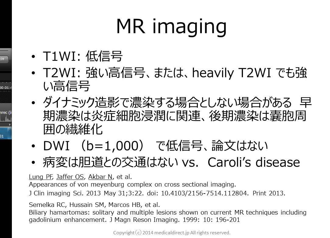
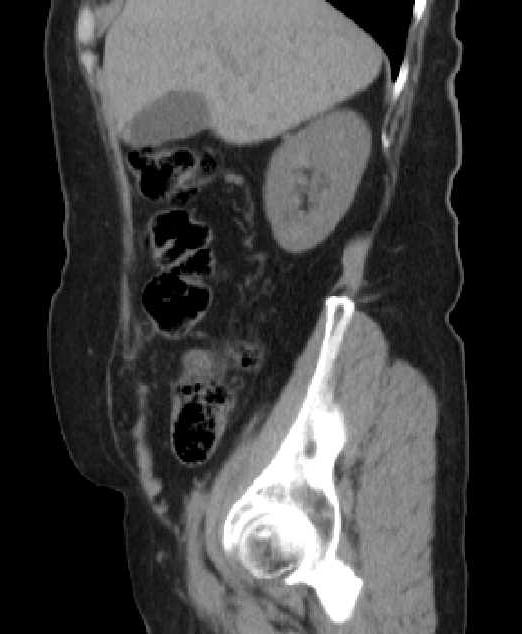
- 半月状線ヘルニア:腹横筋線維が腱膜に移行する腹直筋外縁の半月状線,すなわちSpigel腱膜に発生する稀なヘルニア
- 半月状線の脆弱性、上前腸骨棘を結ぶ線から6cm上方までに発生しやすい=SH belt
- 平均年齢60才台、女性優位
- 左優位、両側性は稀
- 修復術を行った腹壁ヘルニア中の頻度は1%未満
- 原因として外科手術や極度の肥満など
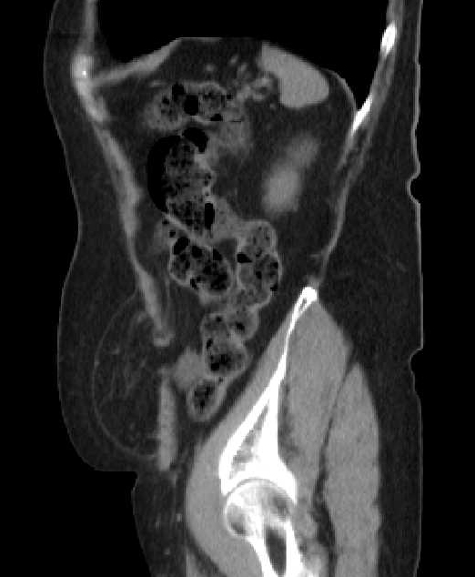
- 外腹斜筋腱膜をこえるか否かで2つに分類
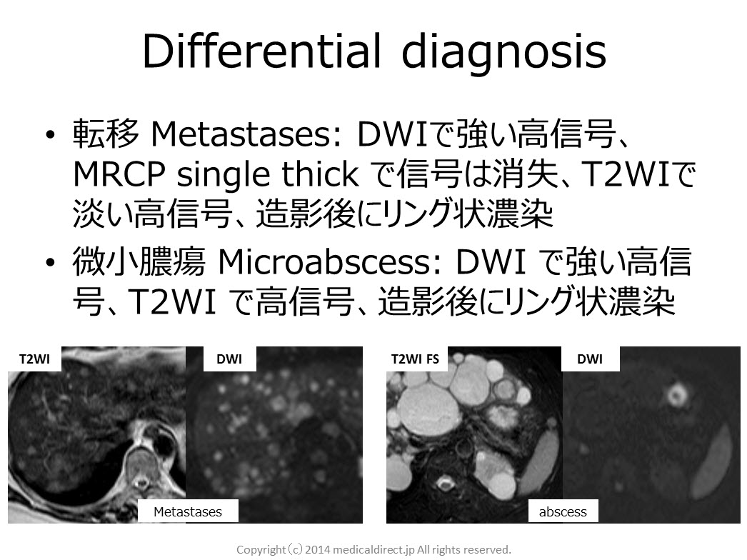
–Interstitial SH: closed loop Small bowel obstruction なり易い
–Subcutaneous SH
- CTで100%の感度とPPV.
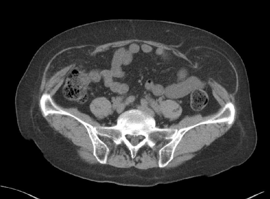
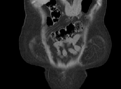
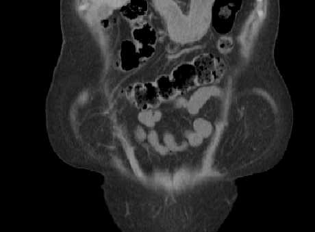
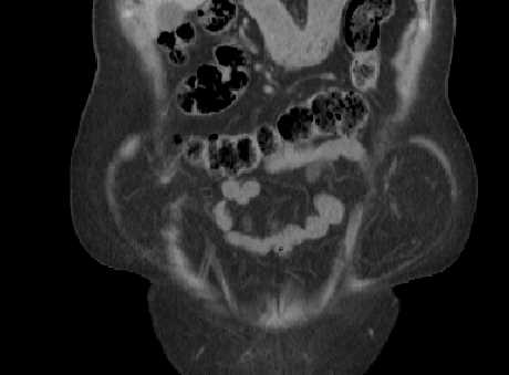
Key images


 文献:
文献:
1.Martine M, Paquette B, Badet N, et al. Spiegelian hernia: CT findings and clinical relevance. Abdom imaging. 2013; 38: 260-264
2. Moles Morenilla L, DocoboDurántez F, Mena Robles J,etal. Spiegelianhernia in Spain. An analysis of 162 cases. Rev EspEnferm Dig. 2005 May;97(5):338-47.
3. Light D, Chattopadhyay D, Bawa S. Radiological and clinical examination in the diagnosis of Spiegelian hernia. Ann R CollSurgEngl 2013; 95:98-10
4. Dabbas N, Adams K, Pearson K, et al. Frequency of abdominal wall hernias: is classical teaching out of date? JRSM Short Rep.2011 Jan 19;2(1):5. doi: 10.1258/shorts.2010.010071.
5. 塩田喜代美、植木孝宣、青井重善、他。CTにて術前診断した半月状線ヘルニアの1例 日臨外会誌 . 2002: 63: 1308-1311
English summary
Summary of Spiegelian hernia
- Spigelian hernias (SH) are rarely reported abdominal wallhernias, which arise from the Spigelianfascia. It lies along the semilunar line lateral to the rectus abdominis muscle.
- SH is due to a weakness of the spiegelian fascia.
- Most of the SH cases occur within the zone, so-called SH belt, which is a transverse band between the line joining both anterior superior iliac spines and parallel line lying 6-cm cranial to it.
- Mean age is 60-70 year-old. Female domination.
- Left side is dominant. Bilateral SH is very rare (2%).
- The incidence of SH among abdominal wall hernia repair surgery is less than 1%.
- Major acquired risk factors of SH are a history of abdominal surgery and obesity.
- Two types of SH are interstitial SH and subcutaneous SH. Closed loop small bowel obstruction is statistically associated with interstitial SH.
- CT has a 100% sensitivity and 100% PPV todiagnose SH.