タグ別アーカイブ: MRI
保護中: 佐志先生の肩を好き version3.0購入の先生のみ イチロウメルマガ特典の特典
保護中: 文献カンファランスシリーズ 第6回(メルマガ購読者のみ再生できます)2013.9.20
保護中: 文献カンファランスシリーズ 第5回(メルマガ購読者のみ再生できます)
保護中: 文献カンファランスシリーズ 第3回(メルマガ購読者のみ再生できます)2012/3/21
Pancreas 症例5 回答に文献、CT, MRI静止画像を追加 2012/3/11
key words: 漿液性嚢胞腺腫、漿液性嚢胞腺癌、膵囊胞性病変
Serous cystadenoma, SCA, SCAs, cystic lesions of the pancreas
漿液性嚢胞腺腫の文献とURL表示
・SCA の病理と対比したMDCTの診断
Shah AA, Sainani NI, Kambadakone AR, et al.
Predictive value of multi-detector computed tomography for accurate diagnosis of serous cystadenoma: radiologic-pathologic correlation.
World J Gastroenterol. 2009 Jun 14;15(22):2739-47.
http://www.ncbi.nlm.nih.gov/pubmed?term=Predictive%20value%20of%20MDCT%20for%20accurate%20diagnosis%20of%20serous
直接PDF(Free)
http://www.wjgnet.com/1007-9327/pdf/v15/i22/2739.pdf
・典型、非典型の漿液性嚢胞腺腫
Choi JY, Kim MJ, Lee JY, et al.
Typical and atypical manifestations of serous cystadenoma of the pancreas: imaging findings with pathologic correlation.
AJR Am J Roentgenol. 2009 Jul;193(1):136-42.
http://www.ncbi.nlm.nih.gov/pubmed/19542405
Free
・漿液性嚢胞腺腫:serous oligocystic adenma とMCTやIPMNとの鑑別の論文
Kim SY, Lee JM, Kim SH, Shin KS, Kim YJ, An SK, Han CJ, Han JK, Choi BI.
Macrocystic neoplasms of the pancreas: CT differentiation of serous oligocystic adenoma from mucinous cystadenoma and intraductal papillary mucinous tumor.
AJR Am J Roentgenol. 2006 Nov;187(5):1192-8.
http://www.ncbi.nlm.nih.gov/pubmed/17056905
Free
・膵嚢胞性病変のまとめ論文
Kalb B, Sarmiento JM, Kooby DA, et al.
MR imaging of cystic lesions of the pancreas.
Radiographics. 2009 Oct;29(6):1749-65.
http://www.ncbi.nlm.nih.gov/pubmed/19959519
2012年3月3日現在Free
・膵充実と囊胞性病変の拡散強調像論文まとめ
Wang Y, Miller FH, Chen ZE, et al.
Diffusion-weighted MR imaging of solid and cystic lesions of the pancreas.
Radiographics. 2011 May-Jun;31(3):E47-64. Review.
http://www.ncbi.nlm.nih.gov/pubmed/21721197
Not Free
・SCN の臨床的特徴の論文
Fukasawa M, Maguchi H, Takahashi K, et al.
Clinical features and natural history of serous cystic neoplasm of the pancreas.
Pancreatology. 2010;10(6):695-701. Epub 2011 Jan 18.
http://www.ncbi.nlm.nih.gov/pubmed/21242709
Free
・PK と SCN, MCT, 偽嚢胞との鑑別
Lv P, Mahyoub R, Lin X, et al.
Differentiating pancreatic ductal adenocarcinoma from pancreatic serous cystadenoma, mucinous cystadenoma, and a pseudocyst with detailed analysis of cystic features on CT scans: a preliminary study.
Korean J Radiol. 2011 Mar-Apr;12(2):187-95. Epub 2011 Mar 3.
http://www.ncbi.nlm.nih.gov/pubmed/21430935
Free
・大腸と脾臓に浸潤した悪性SCCs の症例
Cho W, Cho YB, Jang KT, et al.
Pancreatic serous cystadenocarcinoma with invasive growth into the colon and spleen.
J Korean Surg Soc. 2011 Sep;81(3):221-4. Epub 2011 Sep 26.
http://thesurgery.or.kr/DOIx.php?id=10.4174/jkss.2011.81.3.221
Free
Bano S, Upreti L, Puri SK, Chaudhary V, Sakuja P.
Imaging of pancreatic serous cystadenocarcinoma.
Jpn J Radiol. 2011 Dec;29(10):730-4. Epub 2011 Oct 19.
http://www.ncbi.nlm.nih.gov/pubmed?term=Bano%20S%2C%20Upreti%20L%2C%20Puri%20SK%2C%20Chaudhary%20V%2C%20Sakuja%20P.%20Imaging%20of%20pancreatic%20serous%20cystadenocarcinoma.
Lahat G, Lubezky N, Haim MB, et al.
Cystic tumors of the pancreas: high malignant potential.
Isr Med Assoc J 2011 May;13(5):284-9.
http://www.ima.org.il/imaj/dynamic/web/ArtFromPubmed.asp?year=2011&month=05&page=284
King JC, Ng TT, White SC, Cortina G, Reber HA, Hines OJ.
Pancreatic serous cystadenocarcinoma: a case report and review of the literature.
J Gastrointest Surg. 2009 Oct;13(10):1864-8. Epub 2009 May 21. Review.
http://www.ncbi.nlm.nih.gov/pubmed/19459016
・腫瘍の大きさと部位と臨床所見との関連性
Khashab MA, Shin EJ, Amateau S, et al.
Tumor size and location correlate with behavior of pancreatic serous cystic neoplasms.
Am J Gastroenterol. 2011 Aug;106(8):1521-6. doi: 10.1038/ajg.2011.117. Epub 2011 Apr 5.
http://www.nature.com/ajg/journal/v106/n8/full/ajg2011117a.html
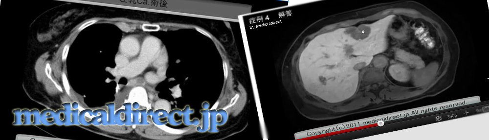
キー画像

膵頭部から鈎部に分葉状の低吸収域腫瘤を認める

膵実質相では強く濃染する充実様に見える部分と囊胞性部分が混在

充実部分にみえ、よく濃染していた部位はwash out されている

DWI では高信号信号を示す

腫瘤はかなり強い高信号を示す。ADC値=2.84×10⁻³mm2/s

MRCP 2D multislce 多数の小嚢胞から構成される

MRCP: Single thick slub 多数の小嚢胞で構成されているT2WI axial 蜂巣状の囊胞性病変
保護中: 文献カンファランスシリーズ 第2回(メルマガ購読者のみ再生できます)
肩関節のMRI: 読影ポイントのすべて
返信

しばらく、ホームページにアップすることが出来ませんでした。あまりの売れ行きにとんでもない値段での販売と成っていたからです。ひどいときには1万7千円程度の中古品が売られていたくらいです。あまりの初版の売れすぎで、全国書店から姿を消していたようです。
当たり前の話しですね。
なぜならば、唯一無二の教科書だからです。日本にはこの教科書しかないのです。だからお勧めというわけではありません。中身もすごいのです。その充実振りたるや、すごいのです。あ、興奮しすぎました。何せ私の肩MRIの師匠の本であるから力が入るのもお無理もありません。
さて、冷静に見ていく事にします。この教科書の今までと異なる点をいくつか列挙します。
1. 執筆者が、放射線科医の独りよがりになっていない点。整形外科医の視点、解剖学の視点からと3つの分野の専門家の共著と成っていることです。このことは、解剖学から入る学生さんから、医者に成り立ての整形外科医、放射線科医はもちろんのこと、放射線専門医となっている私まで広い範囲での読者に受け入れられると言うことです。先ず、実際のページのカウントが始まる前に既に12ページを割いてMRI解剖図譜が載っています。冠状断、矢状断、横断像について細かい解剖が記載されています。しかも、骨については青地で記載されているので筋などとは直ぐに見分けがつくようにされている細やかさです。そして、本格的に解剖の話しに40ページを割いたあと、15ページにわたって肩関節外来での診察について書かれており、放射線科医は整形外科医と会話する場合に必要な知識となるでしょう。その後のメーンイベントである肩関節各論の話しがこれでもかというほど160ペ時にわたって書かれています。最後にMRIの基礎知識がさらに40ページにわたって書かれています。これは、MRIそのものをおびえてしまっている学生さん、整形外科医、若い放射線科医にはもうけものです。そして、これだけでは終わりません。なんと、なんと特典とも言えるような”読影の壺”が43ページにわたり、見開きで一つの疾患をポイントよく説明していく何処かの会社の教科書のような体裁が取られています。メインの160ページのなかに詳細に書かれているにもかかわらずです。
2. 以上書いたように特典を含めた4部構成の305ページ本編と12ページのMRI解剖図譜(推薦の言葉や索引のぞいてです)の合計317ページのコンテンツがこの値段ですのでお買い得としか言いようがありません。つまり、これは、教科書であると同時に辞書なのです。
3.放射線科医が小さい疑問を持っていることに対し、超一流の整形外科医である井樋先生がコメントをするという形の 放射線科医のつぶやき と 整形外科医のコメントは 多の教科書には無い特徴である。例えば、p82の左段での佐志先生のつぶやきは、「ちいさな全層断裂と全層断裂に近い部分断裂とを鑑別することは容易ではありません。これらを鑑別することは臨床的に重要なことでしょうか?」というと 井樋先生からのコメントが「両者は鑑別できなくても問題ありません。部分断裂が深くなってやがて全層断裂になりますから、最後の幕一枚が残っている部分断裂とそこにピンホールの小さい穴があいた全層断裂は臨床的に同一と見なして良いわけです」 と答えておられ、疑問を解消されています。
4.ちょっと息抜きによむコラムの数が半端ではありません。とかく勉強、勉強となりがちな教科書が多い中、ちょっとしたコラムが、こころの清涼剤となることでしょう。コラムには、p61の「断層画像読影の極意」、p88の「腱板はなぜきれるのか?」、p143の「肩甲骨はミズスマシ」、p180の「スポーツは体も心も傷つける」などなど。
先生が得られるメリット:肩関節MRIの全て