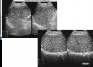
60才台 男性 DM、慢性C型肝炎で内科通院中
サーベイランスにて超音波上肝S6に高輝度SOL指摘
iPad version
You tube version
回答
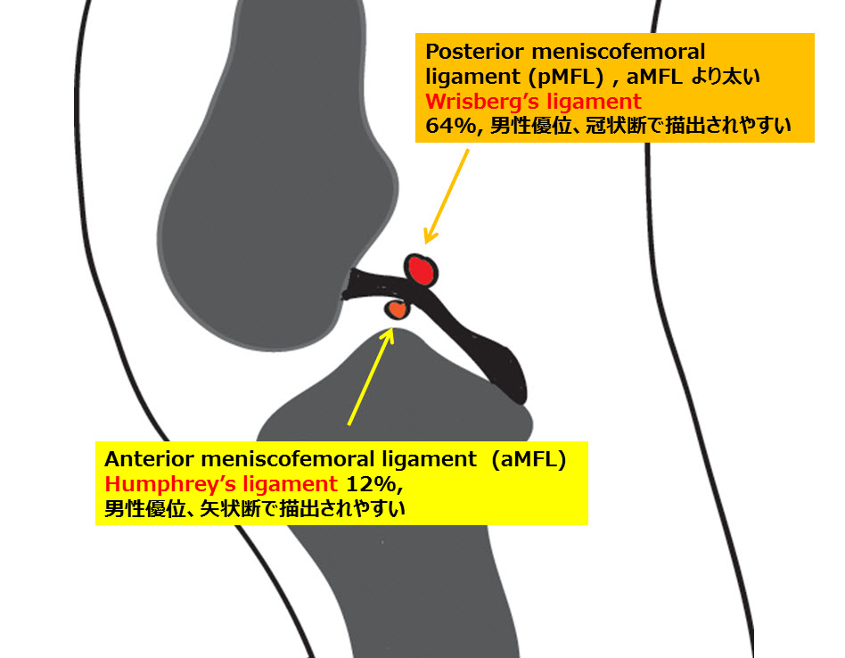
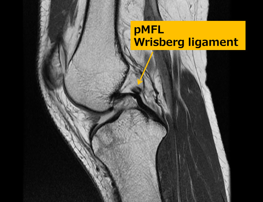
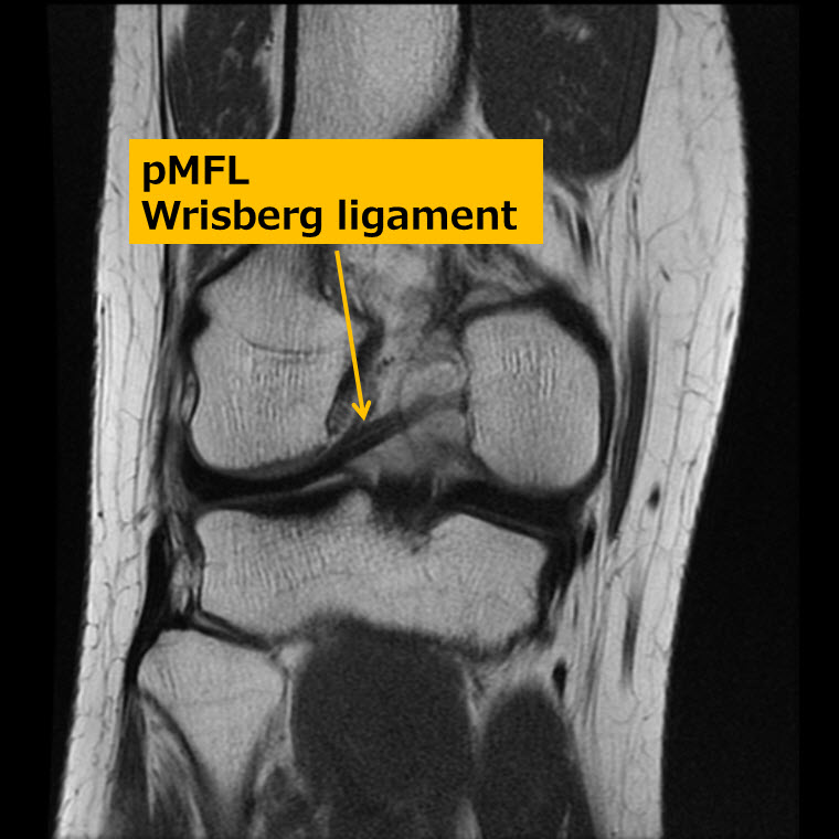
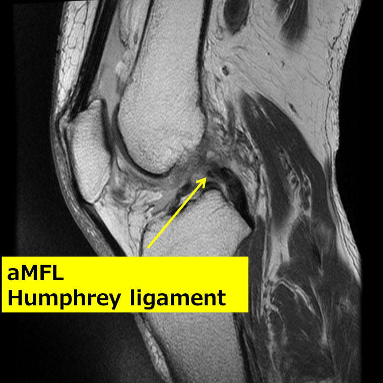
Meniscofemoral ligament (MFL)
iPad version.
You tube version.
Key words: 膝、膝関節、正常構造、ピットフォール、後十字靭帯、半月大腿靭帯、前半月大腿靭帯、後半月大腿靭帯、ハンフリー靭帯、リズバーグ靭帯、knee, knee joint, normal anatomy, pitfall, posterior cruciate ligament, meniscofemoral ligament, anterior meniscofemoral ligament, posterior meniscofemoral ligament, Humphrey’s ligament, Wrisberg’s ligament, Humphrey, Humphry, Wrisberg
Key images: Wrisberg’s ligament 27-year-old man with knee injury.
Wrisberg 靭帯、27歳男性 問題提示症例
Humphrey’s ligament case 82-year-old woman
ハンフリー靭帯 82才女性例
Reference 文献:
1. Bintoudi A1, Natsis K, Tsitouridis I.
Anterior and posterior meniscofemoral ligaments: MRI evaluation.
Anat Res Int. 2012;2012:839724. Epub2012 Sep 17.
–500例中322例 64.4%≒64%
男性240(75%), 女性 82例(25%), 冠状断で描出されやすい
–500例中59例 11.8%≒12%,
男性40(68%), 女性 19例(32%), 矢状断で描出されやすい
2.Watanabe AT, Carter BC, Teitelbaum GP, Bradley WG Jr.
Common pitfalls in magnetic resonance imaging of the knee.
J Bone Joint Surg Am. 1989 Jul;71(6):857-62.
Is the location of the Wrisberg ligament related to frequent complete discoid lateral meniscus tear?
Acta Radiol. 2010 Dec; 51:1120-5. doi: 10.3109/02841851.2010.520026.
4. De Abreu MR, Chung CB, Trudell D, etal.
Skeletal Radiol. 2007;36:729-735. Epub 2007 May 5.
5. Park LS, Jacobson JA, Jamadar DA, et al.
Skeletal Radiol. 2007; 36:399-403. Epub 2007 Jan 16
6. Erbagci H, Yildirim H, Kizilkan N, Gümüsburun E.
An MRI study of the meniscofemoral and transverse ligaments of the knee.
Surg Radiol Anat. 2002 May;24(2):120-4.
解答編
iPad version
You tube version
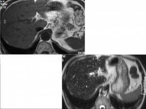
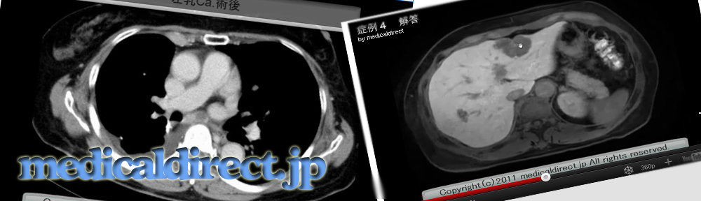
Key words: 副腎癌、副腎腺腫、巨大変性副腎腺腫、Adrenocortical carcinoma, ACC, large degenerated adrenocortical adenoma, pheochromocytoma
Key images.
CT
Plain CT(単純CT)
 Dynamic CT early phase (ダイナミック造影CT 早期相)
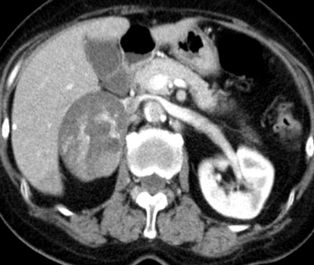
Dynamic CT delayed phase (ダイナミック造影CT後期相)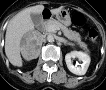 Dynamic CT delayed phase (ダイナミック造影CT後期相): coronal (冠状断)
MRI
T2WI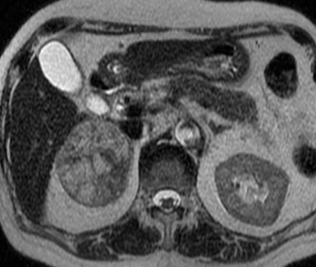 T1WI with opposed phase
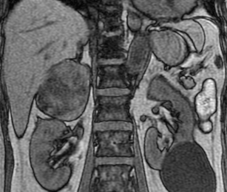 Dynamic MRI early phase
 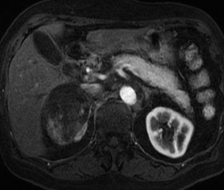 Dynamic MRI portal phase
Dynamic MRI delayed phase, coronal
Diffusion weighted image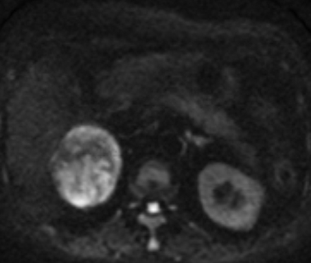
2014/6/16 Adrenal gland adenoma, differentiation, lipid rich adenoma
iphone, iPad の先生は以下
Point words: 0HU, 10HU, 造影前、35秒、5分、15分, Washout, Wash in, 45%, 60% より大, Signal intensity index, 16.5 より大, 10-20HUは MRI, 20HU超えればWashout CT
Key words: adrenal gland tumor, adrenal gland adenoma, lipid rich adenoma, lipid poor adenoma, non adenoma, Washout CT, inphase, opposed phase
Reference:
腺腫 鑑別診断 主要文献
1) Korobkin M, Brodeur FJ, Yutzy GG, ,et al.
AJR Am J Roentgenol. 1996 Mar;166(3):531-6.
http://www.ncbi.nlm.nih.gov/pubmed/8623622
単純CT上のCT値が10HU以下が腺腫の診断の指標と言った古き良き論文
2) Szolar DH, Korobkin M, Reittner P, et al.
Adrenocortical carcinomas and adrenal pheochromocytomas: mass and enhancement loss evaluation at delayed contrast-enhanced CT.
Radiology. 2005 Feb;234(2):479-85.
http://www.ncbi.nlm.nih.gov/pubmed/15671003
3) Singer AA, Obuchowski NA, Einstein DM, et al.
Metastasis or adenoma? Computed tomographic evaluation of the adrenal mass.
Cleve Clin J Med. 1994 May-Jun;61(3):200-5.
http://www.ncbi.nlm.nih.gov/pubmed/?term=Singer%2C+Cleve+Clin+J+Med+1994
Korobkin の前に10HU, 0HUのカットオフ値で検討した論文
4) van Erkel AR, van Gils AP, Lequin M, et al.
CT and MR distinction of adenomas and nonadenomas of the adrenal gland.
J Comput Assist Tomogr.1994 May-Jun;18(3):432-8. http://www.ncbi.nlm.nih.gov/pubmed/8188912
Korobkin の前に10HU, 0HUのカットオフ値で検討した論文
5) Lee MJ, Hahn PF, Papanicolau N, et al.
Benign and malignant adrenal masses: CT distinction with attenuation coefficients, size, and observer analysis.
Radiology. 1991 May;179(2):415-8.
http://www.ncbi.nlm.nih.gov/pubmed/2014283
Korobkin の前に10HU, 0HUのカットオフ値で検討した論文
6) Kamiyama T, Fukukura Y, Yoneyama T, et al.
Distinguishing adrenal adenomas from nonadenomas: combined use of diagnostic parameters of unenhanced and short 5-minute dynamic enhanced CT protocol.
Radiology. 2009 Feb;250(2):474-81. doi: 10.1148/radiol.2502080302. Epub 2008 Nov 26.
http://www.ncbi.nlm.nih.gov/pubmed/19037020
造影前、35秒、5分を用いたWashout CT で診断できるという画期的な論文である。 それまでは15分待つのが常識で、とてもCT室に何もしないで10分以上患者様にいてもらう事はできない。そのためあきらめざるを得なかったWashout CTを現実に運用可能なものとしてくれた。必ずこの論文は取り寄せるべきでしょう。(WO/WI) x100という計算式は同じで、カットオフ値が45%となっている。
7) Park SW, Kim TN, Yoon JH, et al.
The washout rate on the delayed CT image as a diagnostic tool for adrenal adenoma verified by pathology: a multicenter study.
Int Urol Nephrol. 2012 Oct;44(5):1397-402. doi: 10.1007/s11255-012-0202-4. Epub 2012 Jul 14.
http://www.ncbi.nlm.nih.gov/pubmed/22798018
腺腫と非腺腫鑑別のためのWashout CT の “多施設共同研究”
なので、244人の患者と研究の中で抜きんでている患者数
10HUをカットオフ値にすると感度は45%, 特異度は97%
Washout rate を55% で15分の遅延相だと感度94%, 特異度96%
Washout rate を用いる診断法が単純CTでの診断法より優れる
8) Kumagae Y, Fukukura Y Takumi K, et al.
Distinguishing adrenal adenomas from non-adenomas on dynamic enhanced CT: a comparison of 5 and 10 min delays after intravenous contrast medium injection.
Clin Radiol. 2013 Jul;68(7):696-703. doi: 10.1016/j.crad.2013.01.016. Epub 2013 Mar 5.
9) Sangwaiya MJ, Boland GW, Cronin CG, et al.
Incidental adrenal lesions: accuracy of characterization with contrast-enhanced washoutmultidetector CT–10-minute delayed imaging protocol revisited in a large patient cohort.
Radiology. 2010 Aug;256(2):504-10. doi: 10.1148/radiol.10091386.
http://www.ncbi.nlm.nih.gov/pubmed/20656838
323個の副腎腫瘍 をレトロスペクティブに検討だが、単施設ではもっとも検討個数が多い。10分の遅延相はどうなのか? を15分比較して検討したもの。造影前、75秒, 10分 (3ml/s) を使った検討 造影剤は370mgI/ml
10分 でAPWが55% のカットオフ値だと感度62.5%, 特異度93.3%、正診率 64.0% ととにかく15分の時と比較して感度が低くなる カットオフ値60%とするとさらに感度は52%まで低下してしまう。
10) Caoili EM, Korobkin M, Francis IR, et al.
Adrenal masses: characterization with combined unenhanced and delayed enhanced CT.
Radiology. 2002 Mar;222(3):629-33.
http://www.ncbi.nlm.nih.gov/pubmed/11867777
造影前、60秒、15分(2~3ml/s)を使ったデータ APWのカットオフ値は60%としたところ166個の副腎腫瘍 感度98%, 特異度92%, 166この腫瘍中160個(96%)を正しく診断できたという。Korobkin らの教室。
11) Seo JM, Park BK, Park SY, et al.
Characterization of lipid-poor adrenal adenoma: chemical-shift MRI and washout CT.
AJR Am J Roentgenol. 2014 May;202(5):1043-50. doi: 10.2214/AJR.13.11389.
http://www.ncbi.nlm.nih.gov/pubmed/?term=Jung+Min+Seo+Characterization
2014年6月現在の最新の副腎腺腫鑑別についての論文である 必見
Lipid poor adenoma だけを集めたもの Lipid poor adenoma とは単純CTでの腫瘍のCT値が10HUを超えているもの。つまり単純CT上10HUを超えた副腎腫瘍にフォーカスしている点が画期的。そのため、見当数は52個と少ない。その場合にWashout CT と MRIがどちらが信頼できるか? というもので、もちろんCTの方が信頼出来て、腺腫診断の感度はCT が100%、MRIは76%と劣る。特異度もCTは80%あるが、MRIは60%と有意差をもって劣る。しかし、ここからが重要で、実は単純でのCT値が10~20HUの間のLipid poor adenoma 診断についてはCTもMRIも感度は100%と同等。なので、この間の値を単純CT上示した場合は、被爆や造影剤のリスクを考慮すると、第一選択はMRIとなる。ただし、20HUを超える場合は、Washout CTの方がいい。
12) Park BK, Kim CK, Kim B, et al .
Comparison of delayed enhanced CT and chemical shift MR for evaluating hyperattenuating incidental adrenal masses.
Radiology. 2007 Jun;243(3):760-5.
http://www.ncbi.nlm.nih.gov/pubmed/17517932
11) のSeo らと共同演者であるPark BK の同じような単純CTでのCT値が高い腫瘍を集めてMRIの診断能を検討した論文。Washout CTはMRIで診断できない腫瘍の診断が可能であったと言うもの。MRIでの客観指標としてASR と SIIを使用。ASRとはadrenal-to-spleen ratio で SIIはsignal intensity index である。それぞれの計算式は以下
ASR (SIAO/SISO)/(SIAI/SISI) <0.71
SII (SIAI - SIAO) x 100/SIAI > 16.5
13) Outwater EK, Siegelman ES, Huang AB, Birnbaum BA.
Adrenal masses: correlation between CT attenuation value and chemical shift ratio at MR imaging with in-phase and opposed-phase sequences.
Radiology. 1996 Sep;200(3):749-52. Erratum in: Radiology 1996 Dec;201(3):880.
http://www.ncbi.nlm.nih.gov/pubmed/8756926
上記 Signal intensity index の元となっている論文
14) Fujiyoshi F, Nakajo M, Fukukura Y, et al.
Characterization of adrenal tumors by chemical shift fast low-angle shot MR imaging: comparison of four methods of quantitative evaluation.
AJR Am J Roentgenol. 2003 Jun;180(6):1649-57.
http://www.ncbi.nlm.nih.gov/pubmed/12760936
88人102個の副腎腫瘍についての検討なので信頼度は高め
Signal intensity index が最も信頼できる指標であると結論
adrenal-to-spleen ratio, adrenal-to-muscle ratio, adrenal-to-liver ratio と比較
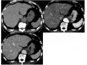
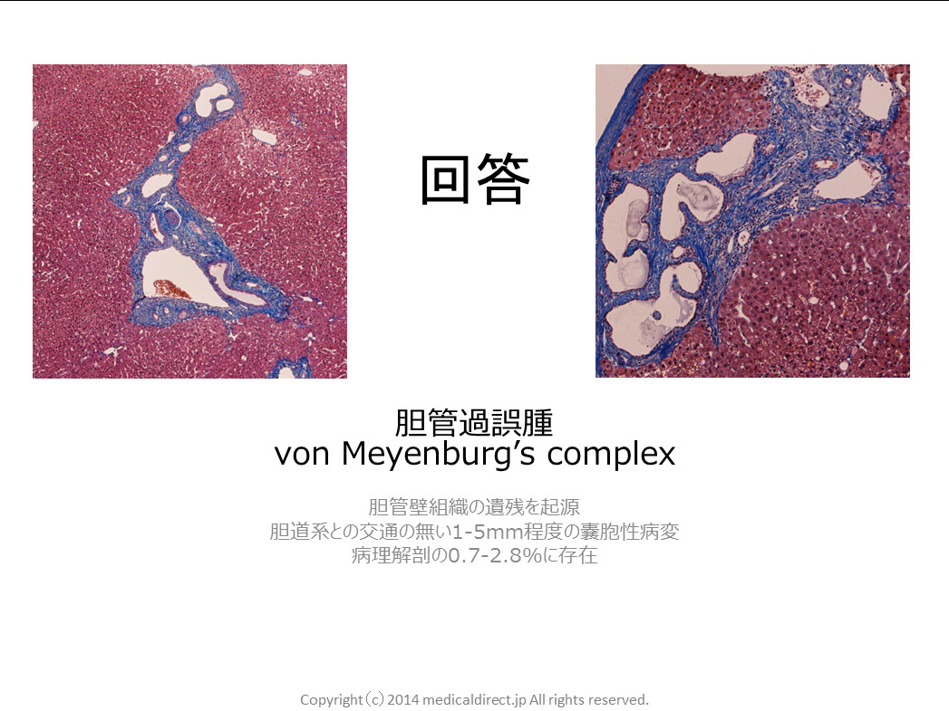
20140407 Multiple hepatic nodules : Answer
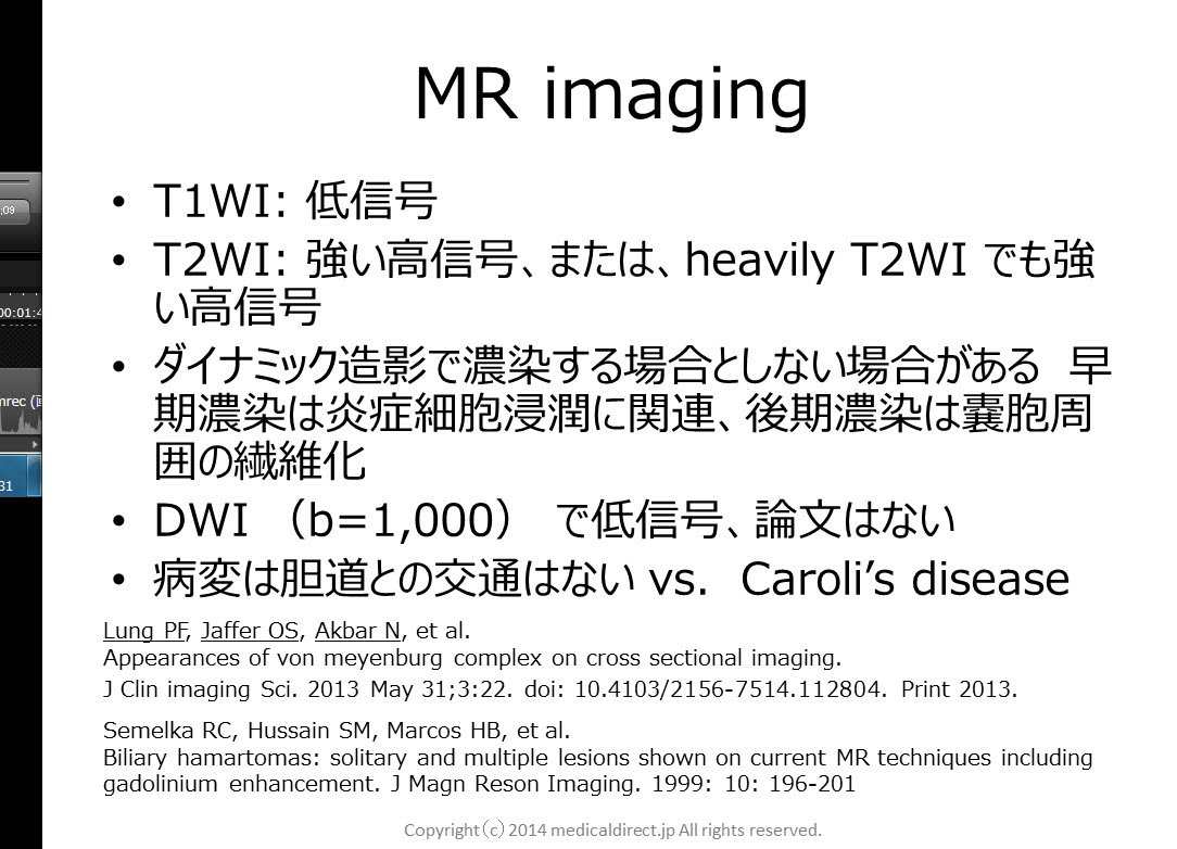
Key words: VMC, von Meyenburg’s complex, biliary hamartoma, 胆管過誤腫、胆管性過誤腫, comet-tail sign, DWI, diffusion weighted imaging, APKD
Key images:
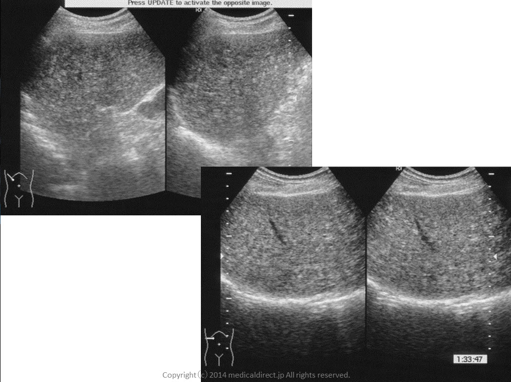
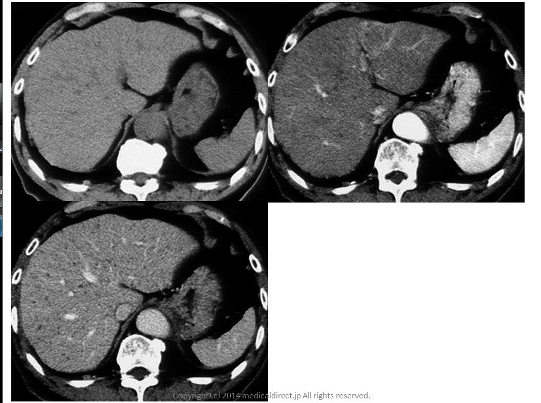
80-year-old man with VMC or biliary hamartoma.

Reference
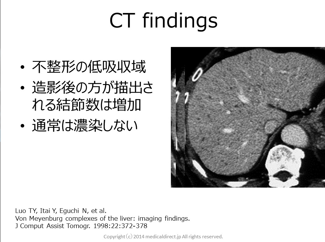
1) Luo TY, Itai Y, Eguchi N, et al.
Von Meyenburg complexes of the liver: imaging findings.
J Comput Assist Tomogr. 1998:22:372-378
2) Lung PF, Jaffer OS, Akbar N,et al.
Appearances of von meyenburg complex on cross sectional imaging.
J Clin imaging Sci. 2013 May 31;3:22. doi: 10.4103/2156-7514.112804. Print 2013.
3) Semelka RC, Hussain SM, Marcos HB, et al.
J MagnReson Imaging. 1999: 10: 196-201
4) MorteléB, Mortelé K, Seynaeve P, et al.
Hepatic bile duct hamartomas (von Meyenburg Complexes): MR and MR cholangiography findings.
J Comput Assist Tomogr. 2002 May-Jun;26(3):438-43.
5) Lev-ToaffAS, Bach AM, Wechsler RJ, et al.
The radiologic and pathologic spectrum of biliary hamartomas.
AJR Am J Roentgenol. 1995 Aug;165(2):309-13.
6) Maher MM, DervanP, Keogh B, et al.
Bile duct hamartomas (von Meyenburg complexes): value of MR imaging in diagnosis.
Abdom Imaging. 1999 Mar-Apr;24(2):171-3.
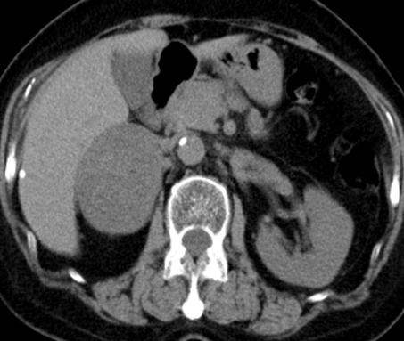
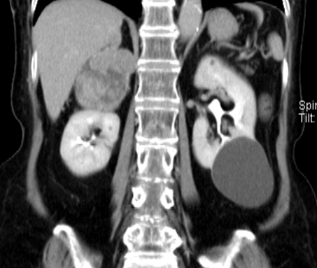
80-year-old man with multiple hepatic nodules.
He has a small gallbladder cancer with 15mm in diameter.
iphone はこちらの動画で