Multiple hepatic nodules 20140527 answer

20140407  Multiple hepatic nodules  : Answer


Key words: VMC, von Meyenburg’s complex, biliary hamartoma,  胆管過誤腫、胆管性過誤腫, comet-tail sign, DWI, diffusion weighted imaging, APKD
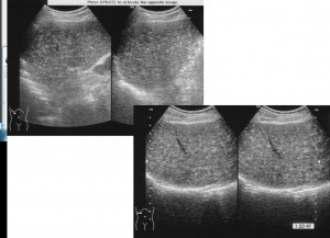
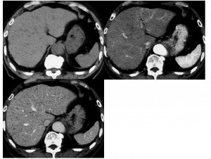
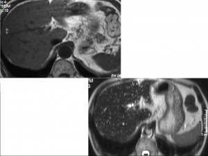
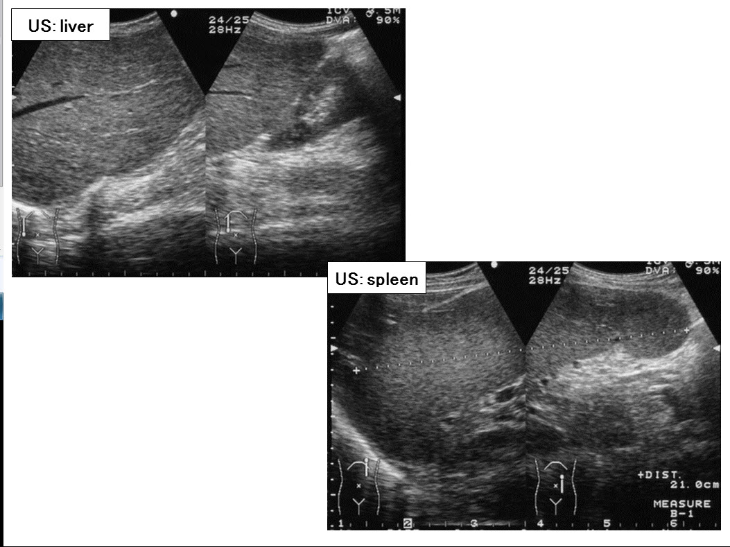
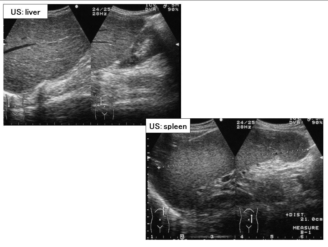
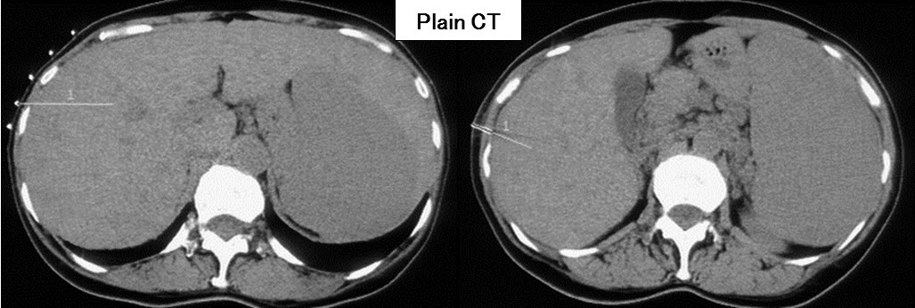
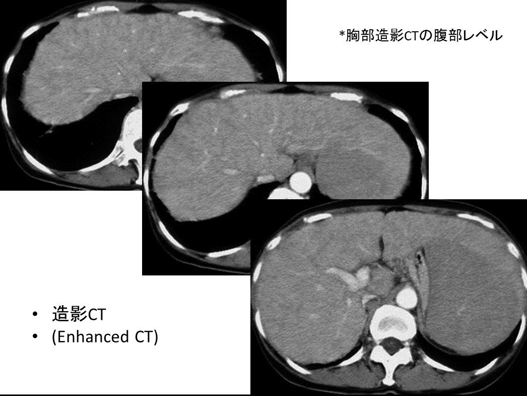
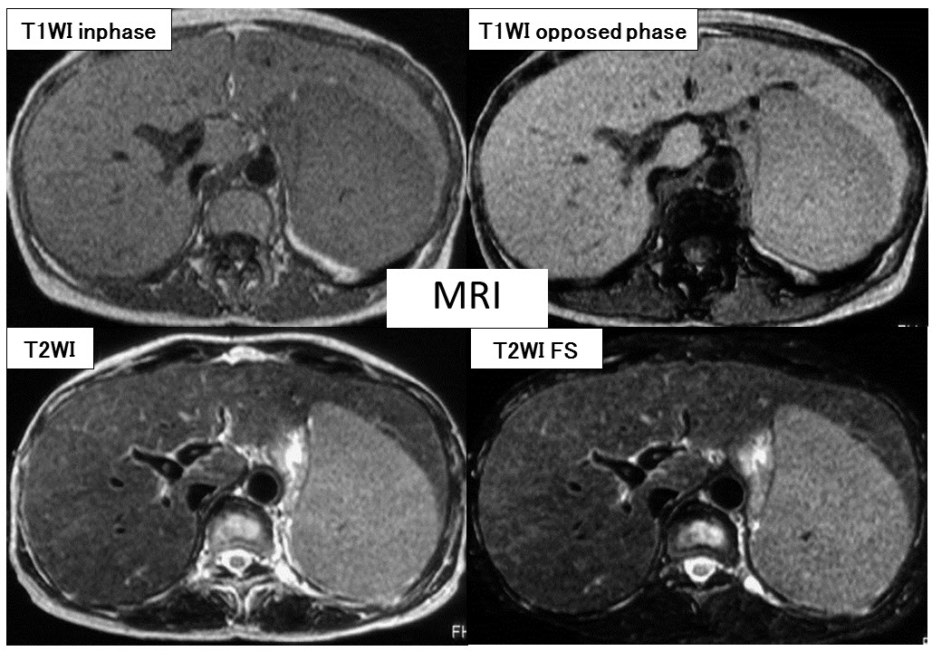
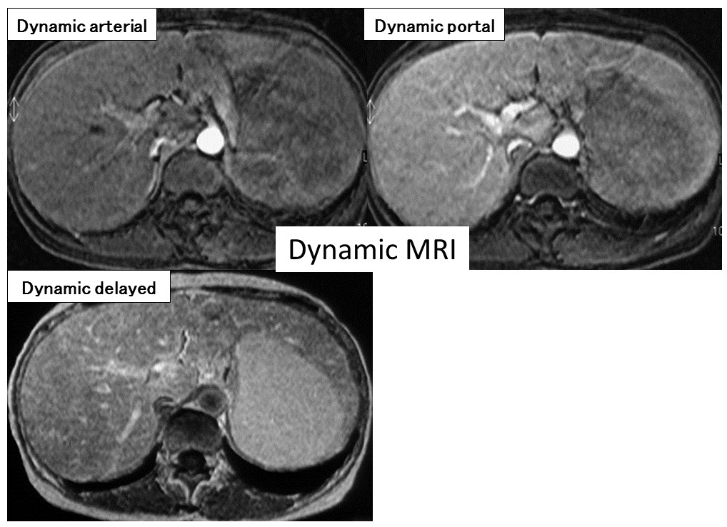
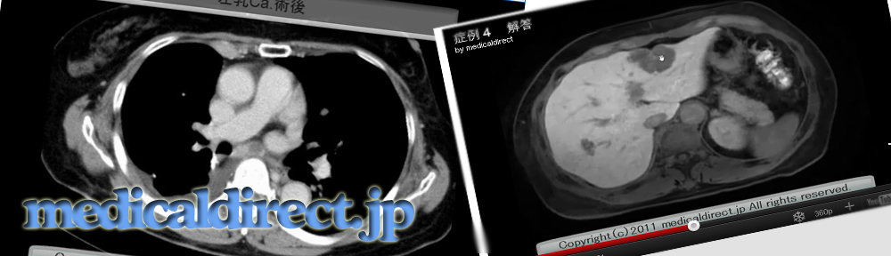
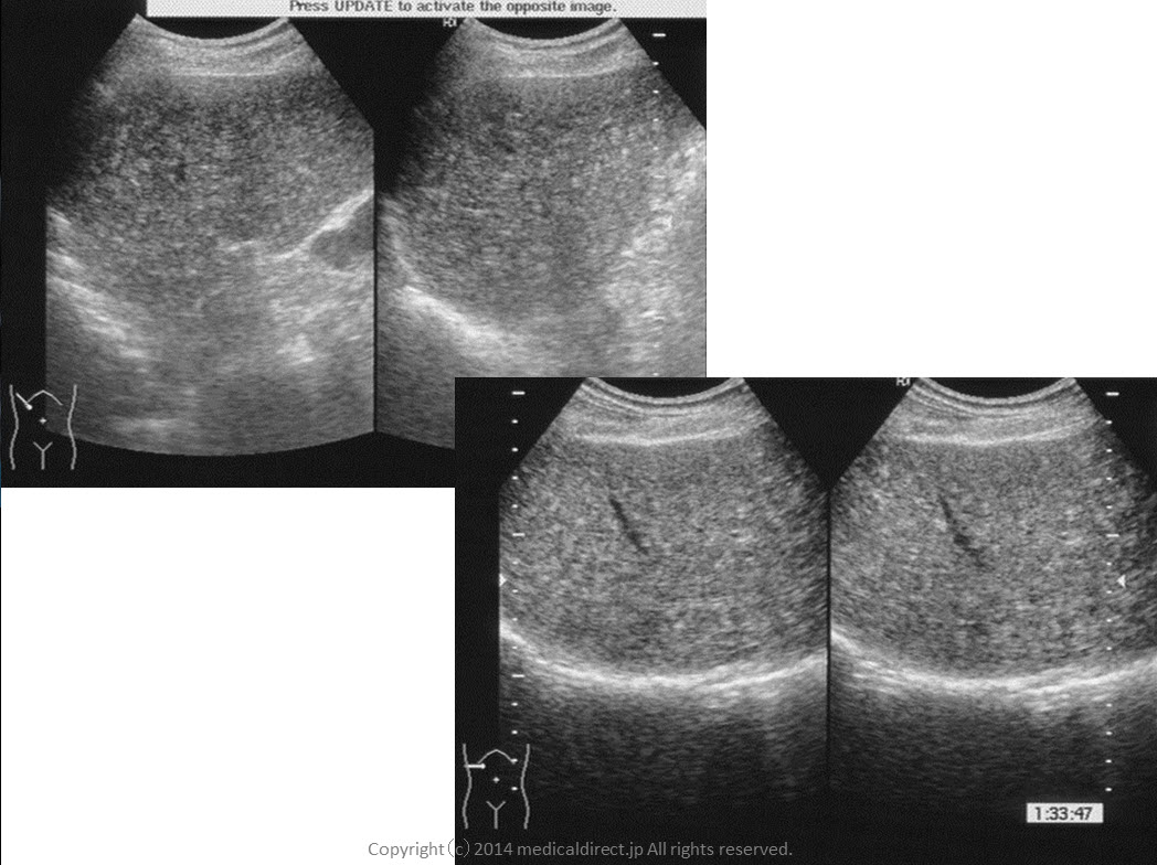
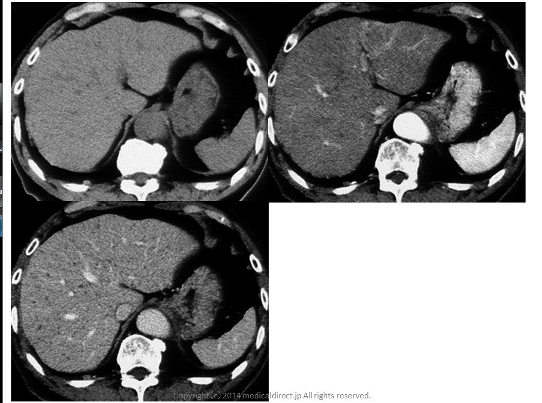
Key images:

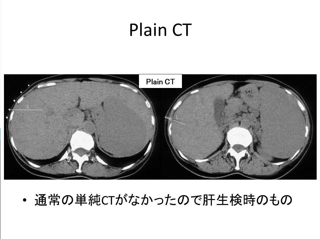
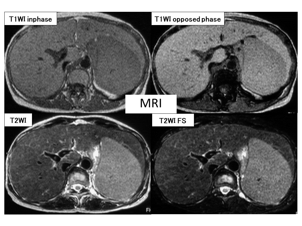
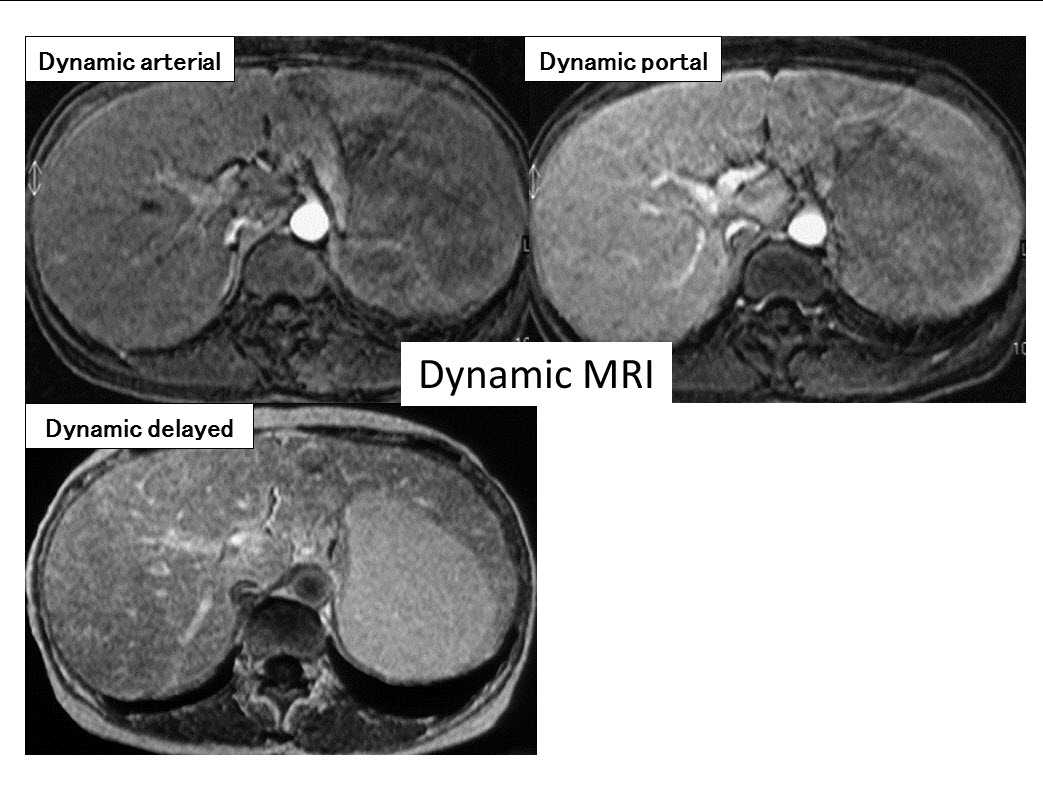
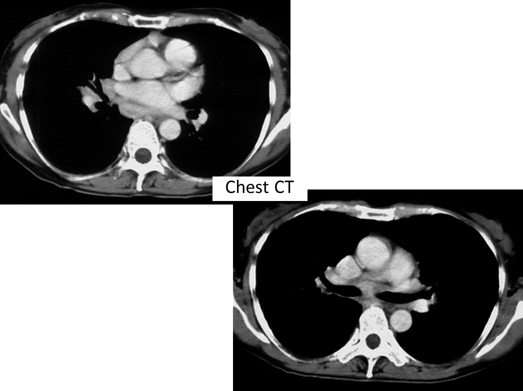
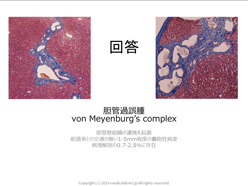
80-year-old man with VMC or biliary hamartoma.


201405 VMC key image US 201405 VMC key image MRI 201405 VMC key image CT 201405 VMC key image Pathology 201405 VMC key image CT findings 201405 VMC key image MRI findings 201405 VMC key image differential diag.


Reference

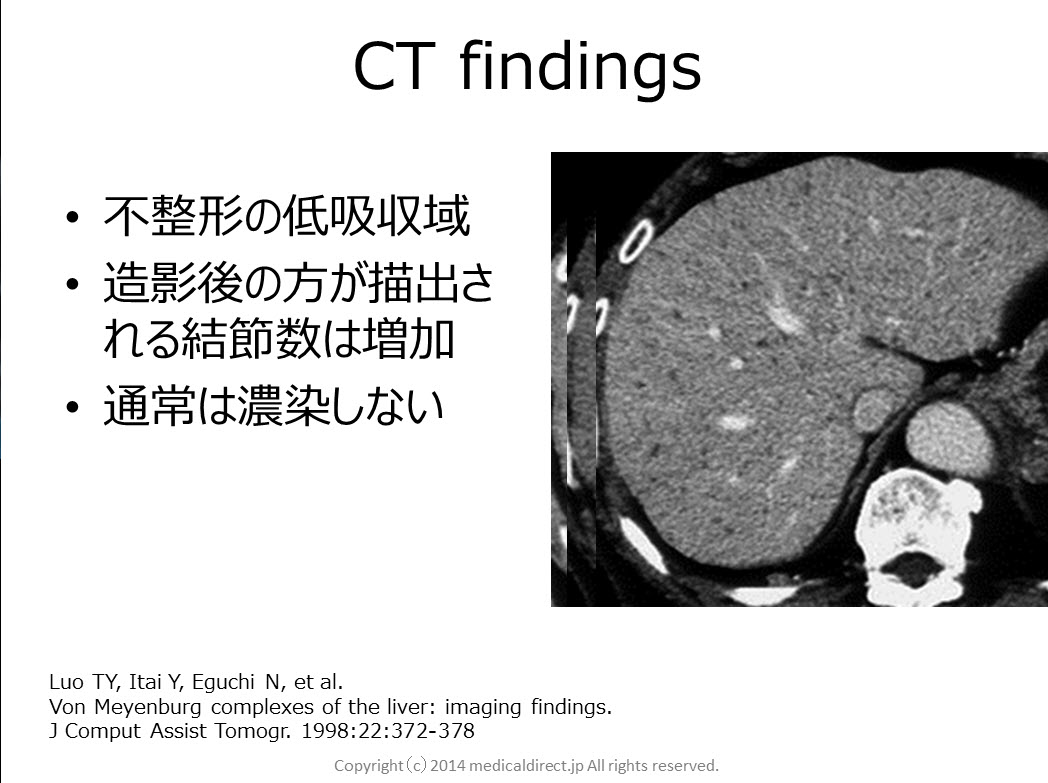
1) Luo TY, Itai Y, Eguchi N, et al.

Von Meyenburg complexes of the liver: imaging findings.

J Comput Assist Tomogr. 1998:22:372-378


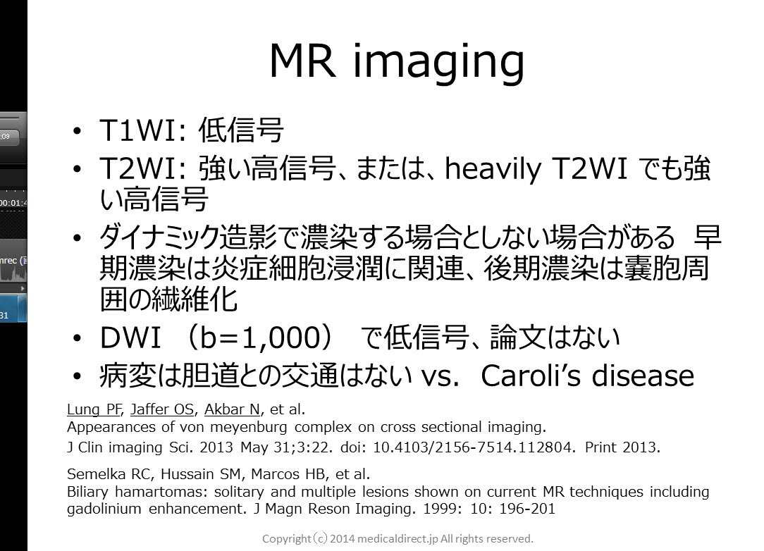
2) Lung PF, Jaffer OS, Akbar N,et al.

Appearances of von meyenburg complex on cross sectional imaging.

J Clin imaging Sci. 2013 May 31;3:22. doi: 10.4103/2156-7514.112804. Print 2013.


3) Semelka RC, Hussain SM, Marcos HB, et al.

Biliary hamartomas: solitary and multiple lesions shown on current MR techniques including gadolinium enhancement.

J MagnReson Imaging. 1999: 10: 196-201


4) MorteléB, Mortelé K, Seynaeve P, et al.

Hepatic bile duct hamartomas (von Meyenburg Complexes): MR and MR cholangiography findings.

J Comput Assist Tomogr.  2002 May-Jun;26(3):438-43.


5)  Lev-ToaffAS, Bach AM, Wechsler RJ, et al.

The radiologic and pathologic spectrum of biliary hamartomas.

AJR Am J Roentgenol.  1995 Aug;165(2):309-13.


6)  Maher MM, DervanP, Keogh B, et al.

Bile duct hamartomas (von Meyenburg complexes): value of MR imaging in diagnosis.

Abdom Imaging. 1999 Mar-Apr;24(2):171-3.


Liver case 13 answer

You tube 動画はこちら

音声のみを聞かれる場合こちら


Key images: クリックすると拡大します


US1Plain CT during biopsyEnhanced CTMRI 1MRI 2Plain film ChestChest CTChest CT 2

Key words:

Sarcoidosis, サルコイドーシス、類上皮非乾落性肉芽腫性疾患、肝脾サルコイドーシス、ACE, CD4/CD8, Panda sign, Hepatic sarcoidosis, Splenic sarcoidosis

Reference:

Warshauer DM, et al. Nodular sarcoidosis of the liver and spleen: analysis of 32 cases. Radiology 1995; 195: 757-762

Elsayes KM, et al. MR imaging of the spleen: spectrum of abnormalities. Radiographics. 2005 ; 25(4):967-82.

 Sakai T, et al. MR imaging of hepatosplenic sarcoidosis. Radiat Med. 1995 Jan-Feb;13(1):39-41.

 Koyama T, et al. Rdiologic manifestations  of sarcoidosis in varisou organs. Radiographics. 2004 24: 87-104

 Warshauer DM, et al. Nodular sarcoidosis of the liver and spleen: appearance on MR images. J Magn Reson Imaging. 1994 Jul-Aug;4(4):553-7.

 Kessler A, et al. Hepatic and splenic sarcoidosis: ultrasound and MR imaging. Abdom Imaging. 1993;18(2):159-63.


Lung case3 回答

iphone ver

key word
Solitary fibrous tumor, SFT, pleura, clinical features

孤立性線維性腫瘍,間葉系細胞由来,臓側胸膜,胸膜,無症状,免疫染色,CD34,粘液変性

Differential diagnosis: pleural mesothelioma 胸膜中皮腫
Other sarcomatous lesions 他の肉腫系病変
すべての年齢,50-60代ピーク,75%が40歳以上
男女差無い,無症状
境界明瞭腫瘍,小さい場合均一,大きくなると不均一
良性はT2WI で低信号
T2WI で高信号の場合良性,悪性の判別難しい
良性でT2WI 高信号となるのは粘液変性,出血,壊死による

Reference 文献
Chu X, Zhang L, Xue Z, et al.

Solitary fibrous tumor of the pleura: An analysis of forty patients.
J Thorac Dis. 2012 Apr 1;4(2):146-54. doi: 10.3978/j.issn.2072-1439.2012.01.05.

Agarwal VK, Plotkin BE, Dumani D, et al.
Solitary fibrous tumor of pleura: a case report and review of clinical, radiographic and histologic findings.
J Radiol Case Rep. 2009;3(5):16-20. doi: 10.3941/jrcr.v3i5.200. Epub 2009 May 1.

Sekiya M, Yoshimi K, Muraki K, et al.
Solitary fibrous tumor of the pleura: ultrasonographic imaging findings of 3 cases.
Respir Investig. 2013 Sep;51(3):200-4. doi: 10.1016/j.resinv.2013.04.001. Epub 2013 Jun 13.

Moureau-Zabotto L, Chetaille B, et al.
Solitary fibrous tumor of the prostate: case report and review of the literature.
Case Rep Oncol. 2012 Jan;5(1):22-9. doi: 10.1159/000335680. Epub 2012 Jan 10.

Li JP, Xie CM, Zhang R,et al.
[Imaging features and clinicopathological manifestations of solitary fibrous tumors].
Zhonghua Zhong Liu Za Zhi. 2010 May;32(5):363-7. Chinese.

Nomura T, Satoh R, Kashima K, et al.
A case of large solitary fibrous tumor in the retroperitoneum.
Clin Med Case Rep. 2009 Mar 23;2:21-5. eCollection 2009.

Rosado-de-Christenson ML, Abbott GF, McAdams HP, et al.
From the archives of the AFIP: Localized fibrous tumor of the pleura.
Radiographics. 2003 May-Jun;23(3):759-83.

Inaoka T, Takahashi K, Miyokawa N, et al.
Solitary fibrous tumor of the pleura: apparent diffusion coefficient (ADC) value and ADC map to predict malignant transformation.
J Magn Reson Imaging. 2007 Jul;26(1):155-8.