↓iphone用動画
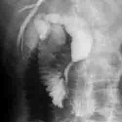
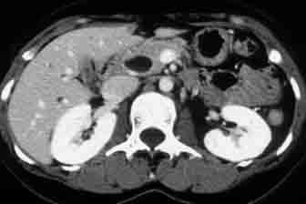
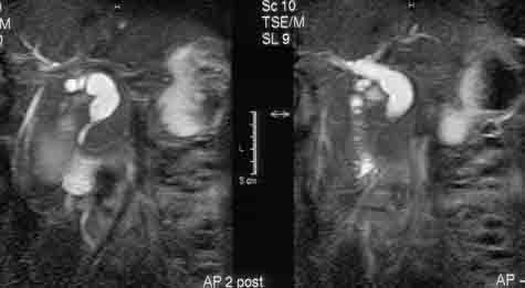
症例3の回答 27才女性 主訴:繰り返す腹痛
Key words: 総胆管嚢腫、膵胆管合流異常症、MRCP, MR cholangiography, MR cholangiopancreatography, ERCP, Todani分類、Todani’s classification, 胆管癌、膵炎
文献:
・Lam WW, Lam, et al. MR cholangiography and CT cholangiography of pediatric patients with choledochal cysts. AJR Am J Roentgenol. 1999 ;173:401-5.
http://www.ajronline.org/cgi/reprint/173/2/401
・Kim OH, et al. Imaging of the choledochal cyst. Radiographics 1995;15:69-88
http://radiographics.rsna.org/content/15/1/69.long
・Vitellas KM, et al. MR Cholangiopancreatography of bile and pancreatic duct abnormalities with emphasis on the single-shot fast spin-echo technique. Radiographics 2000; 20:939?957
http://radiographics.rsna.org/content/20/4/939.long
・Irie H, et al. Value of MR cholangiopancreatography in evaluating choledochal cysts. AJR Am J Roentgenol. 1998;171:1381-1385
http://www.ajronline.org/cgi/reprint/171/5/1381
<key image>
ERCP

US

enhancedCT

MRCP

症例2の回答 55歳男性 体重減少、心窩部痛、1ヶ月前頃より上腹部のつかえ感あり腹部膨満感自覚。5日で5キロの体重減少あり
Key words: AIP, autoimmune pancreatitis, 自己免疫性膵炎、IgG4関連疾患、IgG4-related sclerosing disease, IgG4-related sclerosing cholangitis, IgG4関連硬化性胆管炎、IgG4-related sclerosing sialadenitis, IgG4関連唾液腺炎、Mikulicz diasese, ミクリッツ病, IgG4, steroid, ステロイド
参考文献
・Kamisawa T, et al. Autoimmune pancreatitis: proposal of IgG4-related sclerosing disease. J Gastroenterol 2006; 41:613-625
・Sahani DV, et al. Autoimmune pancreatitis: Imaging features. Radiology 2004; 233:345-352
・Ghazale A, et al. Value of serum IgG4 in the diagnosis of autoimmune pancreatitis and in distinguishing it from pancreatic cancer. Am J Gastroenterol. 2007; 102: 1646-1653
・Manfredi R, et al. Autoimmune pancreatitis: CT patterns and their changes after steroid treatment. Radiology 247; 435-443
http://radiology.rsna.org/content/247/2/435.full.pdf+html
・Kawamoto S, et al. Lymphoplasmacytic Sclerosing Pancreatitis with Obstructive Jaundice: CT and Pathology Features. AJR 2004;183:915-921
http://www.ajronline.org/cgi/reprint/183/4/915
・Kamisawa T, et al. IgG4-related sclerosing disease. World J Gastroenterol. 2008; 14: 3948-3955
http://www.wjgnet.com/1007-9327/14/3948.pdf
・Kamisawa T, et al. Sclerosing cholangitis associated with autoimmune pancreatitis differs from primary sclerosing cholangitis. World J Gastroenterol 2009; 15:2357-2360
http://www.wjgnet.com/1007-9327/15/2357.pdf
・Taguchi M, et al. Autoimmune pancreatitis with IgG4-positive plasma cell infiltration in salivary glands and biliary tract. World J Gastroenterol 2005; 11:5577-5581
・Irie H, et al. Autoimmune pancreatitis: CT and MR characteristics. AJR 1998; 170:1323-1327
http://www.ajronline.org/cgi/reprint/170/5/1323
・Takahashi N, et al. Renal involvement in patients with autoimmune pancreatitis: CT and MR imaging findings. Radiology 2007; 242:791-801
http://radiology.rsna.org/content/242/3/791.full.pdf+html
・Park SJ, et al. Clinical characteristics, recurrence features, and treatment outcomes 55 patients with autoimmune pancreatitis. Korean J Gastroenterl. 2008;52:230-246
・Suga K, et al. F-18 FDG PET-CT findings in Mikulicz diseaase and systemic involvement of IgG4-related lesions. Clin Nucl Med. 2009; 34:164-167
・Kim KP, et al. Autoimmune chronic pancreatitis. Am J Gastroenterol 2004;99:1605-1616
・Park SJ, et al. Clinical characteristics, recurrence features, and treatment outcomes 55 patients with autoimmune pancreatitis. Korean J Gastroenterl. 2008;52:230-246
・Suga K, et al. F-18 FDG PET-CT findings in Mikulicz diseaase and systemic involvement of IgG4-related lesions. Clin Nucl Med. 2009; 34:164-167
・Taniguchi T, et al. Diffusion-weighted magnetic resonance imaging in autoimmune pancreatitis. Jpn J Radiol. 2009;27:138-42. Epub 2009 May 3.
・Takahashi N, et al. Autoimmune pancreatitis: differentiation from pancreatic carcinom and normal pancreas on the basis of enhancement characteristics at dual-phase CT. AJR 2009;193:479-484
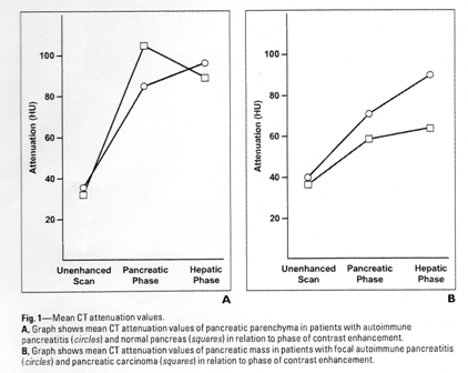
この文献より限局型PKとAIP鑑別のためのダイナミック造影パターンのFig1の図を以下に引用。

上記左図Aは、正常膵とAIPのダイナミックパターンの比較
四角が正常、丸がAIPです。
四角の正常に比較してAIPは膵実質相の濃染が悪く
肝実質相(門脈相)では両者に濃染の差はありません。
上記右図Bは、PKとAIPのダイナミックパターンの比較
丸がAIP、四角がPKです。
AIPは肝実質相にかけて濃染が高まりますが、
PKは肝実質相でも濃染不良となっています。
つまり、一言でいえば、AIPは、
「膵実質相では濃染が遅延するが肝実質相では正常と変わらない」
となります。
by イチロウ の補足解説でした。
Key Words::成熟奇形腫破裂, ruptured teratoma,石灰化、歯、毛髪、脂肪,急性腹膜炎、慢性肉芽腫性腹膜炎
文献:
Park SB, et al. Imaging findings of complications and unusual manifestations of ovarian teratomas. Radiographics. 2008;28:969-83
http://radiographics.rsna.org/content/28/4/969.full.pdf+html
Fibus TF Intraperitoneal rupture of a benign cystic ovarian teratoma: findings at CT and MR imaging. AJR Am J Roentgenol. 2000;174:261-2
http://www.ajronline.org/cgi/reprint/174/1/261
Rha SE, Byun JY, Jung SE, et al. Atypical CT and MRI manifestations of mature ovarian cystic teratomas.?AJR Am J Roentgenol 2004;183:743?750.
http://www.ajronline.org/cgi/reprint/183/3/743