作成者アーカイブ: イチロウ
保護中: 佐志先生の肩を好き version3.0購入の先生のみ イチロウメルマガ特典の特典
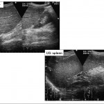
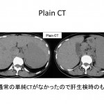
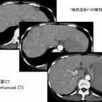
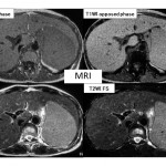
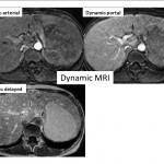
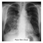
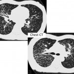
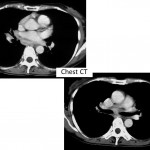
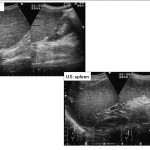
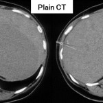
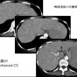
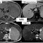
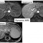
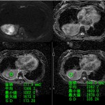
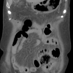
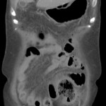
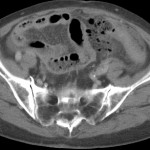
Liver case 13 answer
You tube 動画はこちら
音声のみを聞かれる場合こちら
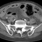
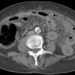
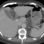
Key images: クリックすると拡大します
Key words:
Sarcoidosis, サルコイドーシス、類上皮非乾落性肉芽腫性疾患、肝脾サルコイドーシス、ACE, CD4/CD8, Panda sign, Hepatic sarcoidosis, Splenic sarcoidosis
Reference:
Warshauer DM, et al. Nodular sarcoidosis of the liver and spleen: analysis of 32 cases. Radiology 1995; 195: 757-762
Elsayes KM, et al. MR imaging of the spleen: spectrum of abnormalities. Radiographics. 2005 ; 25(4):967-82.
Sakai T, et al. MR imaging of hepatosplenic sarcoidosis. Radiat Med. 1995 Jan-Feb;13(1):39-41.
Koyama T, et al. Rdiologic manifestations of sarcoidosis in varisou organs. Radiographics. 2004 24: 87-104
Warshauer DM, et al. Nodular sarcoidosis of the liver and spleen: appearance on MR images. J Magn Reson Imaging. 1994 Jul-Aug;4(4):553-7.
Kessler A, et al. Hepatic and splenic sarcoidosis: ultrasound and MR imaging. Abdom Imaging. 1993;18(2):159-63.
Liver case13 Question
Lung case3 回答
iphone ver
key word
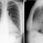
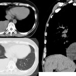
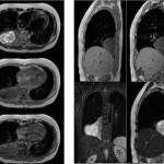
Solitary fibrous tumor, SFT, pleura, clinical features
孤立性線維性腫瘍,間葉系細胞由来,臓側胸膜,胸膜,無症状,免疫染色,CD34,粘液変性
Differential diagnosis: pleural mesothelioma 胸膜中皮腫
Other sarcomatous lesions 他の肉腫系病変
すべての年齢,50-60代ピーク,75%が40歳以上
男女差無い,無症状
境界明瞭腫瘍,小さい場合均一,大きくなると不均一
良性はT2WI で低信号
T2WI で高信号の場合良性,悪性の判別難しい
良性でT2WI 高信号となるのは粘液変性,出血,壊死による
Reference 文献
Chu X, Zhang L, Xue Z, et al.
Solitary fibrous tumor of the pleura: An analysis of forty patients.
J Thorac Dis. 2012 Apr 1;4(2):146-54. doi: 10.3978/j.issn.2072-1439.2012.01.05.
Agarwal VK, Plotkin BE, Dumani D, et al.
Solitary fibrous tumor of pleura: a case report and review of clinical, radiographic and histologic findings.
J Radiol Case Rep. 2009;3(5):16-20. doi: 10.3941/jrcr.v3i5.200. Epub 2009 May 1.
Sekiya M, Yoshimi K, Muraki K, et al.
Solitary fibrous tumor of the pleura: ultrasonographic imaging findings of 3 cases.
Respir Investig. 2013 Sep;51(3):200-4. doi: 10.1016/j.resinv.2013.04.001. Epub 2013 Jun 13.
Moureau-Zabotto L, Chetaille B, et al.
Solitary fibrous tumor of the prostate: case report and review of the literature.
Case Rep Oncol. 2012 Jan;5(1):22-9. doi: 10.1159/000335680. Epub 2012 Jan 10.
Li JP, Xie CM, Zhang R,et al.
[Imaging features and clinicopathological manifestations of solitary fibrous tumors].
Zhonghua Zhong Liu Za Zhi. 2010 May;32(5):363-7. Chinese.
Nomura T, Satoh R, Kashima K, et al.
A case of large solitary fibrous tumor in the retroperitoneum.
Clin Med Case Rep. 2009 Mar 23;2:21-5. eCollection 2009.
Rosado-de-Christenson ML, Abbott GF, McAdams HP, et al.
From the archives of the AFIP: Localized fibrous tumor of the pleura.
Radiographics. 2003 May-Jun;23(3):759-83.
Inaoka T, Takahashi K, Miyokawa N, et al.
Solitary fibrous tumor of the pleura: apparent diffusion coefficient (ADC) value and ADC map to predict malignant transformation.
J Magn Reson Imaging. 2007 Jul;26(1):155-8.

