・Lam WW, Lam, et al. MR cholangiography and CT cholangiography of pediatric patients with choledochal cysts. AJR Am J Roentgenol. 1999 ;173:401-5. http://www.ajronline.org/cgi/reprint/173/2/401
・Vitellas KM, et al. MR Cholangiopancreatography of bile and pancreatic duct abnormalities with emphasis on the single-shot fast spin-echo technique. Radiographics 2000; 20:939?957 http://radiographics.rsna.org/content/20/4/939.long
・Kamisawa T, et al. Autoimmune pancreatitis: proposal of IgG4-related sclerosing disease. J Gastroenterol 2006; 41:613-625
・Sahani DV, et al. Autoimmune pancreatitis: Imaging features. Radiology 2004; 233:345-352
・Ghazale A, et al. Value of serum IgG4 in the diagnosis of autoimmune pancreatitis and in distinguishing it from pancreatic cancer. Am J Gastroenterol. 2007; 102: 1646-1653
・Manfredi R, et al. Autoimmune pancreatitis: CT patterns and their changes after steroid treatment. Radiology 247; 435-443 http://radiology.rsna.org/content/247/2/435.full.pdf+html
・Kawamoto S, et al. Lymphoplasmacytic Sclerosing Pancreatitis with Obstructive Jaundice: CT and Pathology Features. AJR 2004;183:915-921 http://www.ajronline.org/cgi/reprint/183/4/915
・Kamisawa T, et al. IgG4-related sclerosing disease. World J Gastroenterol. 2008; 14: 3948-3955 http://www.wjgnet.com/1007-9327/14/3948.pdf
・Kamisawa T, et al. Sclerosing cholangitis associated with autoimmune pancreatitis differs from primary sclerosing cholangitis. World J Gastroenterol 2009; 15:2357-2360 http://www.wjgnet.com/1007-9327/15/2357.pdf
・Taguchi M, et al. Autoimmune pancreatitis with IgG4-positive plasma cell infiltration in salivary glands and biliary tract. World J Gastroenterol 2005; 11:5577-5581
・Irie H, et al. Autoimmune pancreatitis: CT and MR characteristics. AJR 1998; 170:1323-1327 http://www.ajronline.org/cgi/reprint/170/5/1323
・Takahashi N, et al. Renal involvement in patients with autoimmune pancreatitis: CT and MR imaging findings. Radiology 2007; 242:791-801 http://radiology.rsna.org/content/242/3/791.full.pdf+html
・Park SJ, et al. Clinical characteristics, recurrence features, and treatment outcomes 55 patients with autoimmune pancreatitis. Korean J Gastroenterl. 2008;52:230-246
・Suga K, et al. F-18 FDG PET-CT findings in Mikulicz diseaase and systemic involvement of IgG4-related lesions. Clin Nucl Med. 2009; 34:164-167
・Kim KP, et al. Autoimmune chronic pancreatitis. Am J Gastroenterol 2004;99:1605-1616
・Park SJ, et al. Clinical characteristics, recurrence features, and treatment outcomes 55 patients with autoimmune pancreatitis. Korean J Gastroenterl. 2008;52:230-246
・Suga K, et al. F-18 FDG PET-CT findings in Mikulicz diseaase and systemic involvement of IgG4-related lesions. Clin Nucl Med. 2009; 34:164-167
・Taniguchi T, et al. Diffusion-weighted magnetic resonance imaging in autoimmune pancreatitis. Jpn J Radiol. 2009;27:138-42. Epub 2009 May 3.
・Takahashi N, et al. Autoimmune pancreatitis: differentiation from pancreatic carcinom and normal pancreas on the basis of enhancement characteristics at dual-phase CT. AJR 2009;193:479-484
Key words: 巨大膵腫瘍、進行早い膵癌、膵癌、退形成性癌、膵巨細細胞型、破骨細胞型、Anaplastic carcinoma of pancreas、GCT of pancreas, giant cell carcinoma of osteoclastoid type, Pleomorphic carcinoma
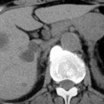
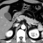
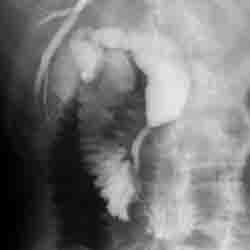
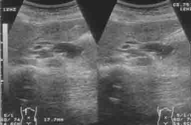
Key images:
退形成性膵癌を疑う時はどんな時?
①初診時に既にかなり進行した膵および膵周囲腫瘤
②膵癌が疑われるが、腫瘍マーカー上昇がない、白血球上昇がある
③巨大な充実性腫瘍で、内部壊死傾向強い
References:
1. Nakajima A, et al. Anaplastic carcinoma of the pancreas producing granulocyte-colony stimulating factor: a case report. J Med Case Reports. 2008;2:391
2. Oyama Y, et al. Relatively rare Osteoclast-type giant cell tumor of the pancreas-resection case. Fukubuhoushasennkenkyukai. 2003;9:126-127
3. Nojima T et al. Pleomorphic carcinoma of the pancreas with osteoclast-like giant cells. Int J Pancreatol. 1993;14275-281
4. Ichikawa T, et al. Atypical exocrine and endocrine pancreatic tumors (anaplastic, small cell, and giant cell types): CT and pathologic features in 14 patients. Abdom Imaging. 2000; 25:409-419 論文リンクはこちら
1. Sano M, Homma T, Hayashi E, et al. Clinicopathological characteristics of anaplastic carcinoma of the pancreas with rhabdoid features. Virchows Arch. 2014 Nov;465(5):531-8. doi: 10.1007/s00428-014-1631-5. Epub 2014 Jul 17. リンクはこちら
2. Jones TS, Jones EL, McManus M, et al. Multifocal anaplasticpancreatic carcinoma requiring neoadjuvant chemotherapy and total pancreatectomy: report of a case. JOP. 2013 May 10;14(3):289-91. doi: 10.6092/1590-8577/1416.
3. Sanada Y, Yoshida K, Itoh M, Okita R, Okada M. Invasive ductal carcinoma of the pancreas showing exophytic growth. Hepatobiliary Pancreat Dis Int. 2009 Feb;8(1):97-102.] 論文リンクはこちら
4. Nakajima A, Takahashi H, Inamori M, et al. Anaplastic carcinoma of the pancreas producing granulocyte-colony stimulating factor: a case report. J Med Case Rep. 2008 Dec 17;2:391. doi: 10.1186/1752-1947-2-391. 論文リンクはこちら
1. Hammond NA, Miller FH, Day K, et al. Imaging features of the less common pancreatic masses. Abdom Imaging. 2013 Jun;38(3):561-72. doi: 10.1007/s00261-012-9922-2. Review. 画像診断という点では 一番最新論文か 論文リンクはこちら
2. Gotohda N, Nakagohri T, Saito N, et al. A case of anaplastic ductal carcinoma of the pancreas with production of granulocyte-colony stimulating factor. Hepatogastroenterology. 2006 Nov-Dec;53(72):957-9.
3. Ichikawa T, Federle MP, Ohba S, et al. Atypical exocrine and endocrine pancreatic tumors (anaplastic, small cell, and giant cell types): CT and pathologic features in 14 patients. Abdom Imaging. 2000 Jul-Aug;25(4):409-19. 上記既に記載済み(重複)
4. Okazaki M, Makino I, Kitagawa H, et al. A case report of anaplastic carcinoma of the pancreas with remarkable intraductal tumor growth into the main pancreatic duct. World J Gastroenterol. 2014 Jan 21;20(3):852-6. doi: 10.3748/wjg.v20.i3.852
5. Zou XP, Yu ZL, Li ZS, Zhou GZ. Clinicopathological features of giant cell carcinoma of thepancreas. Hepatobiliary Pancreat Dis Int. 2004 May;3(2):300-2.